 Explore
 Validate
 Learn
Learn Western blot
 Immunocytochemistry
Antibody data
- Antibody Data
- Antigen structure
- References [13]
- Comments [0]
- Validations
- Western blot [6]
- Immunohistochemistry [1]
- Other assay [1]
Submit
Validation data
Reference
Comment
Report error
- Product number
- NB100-654 - Provider product page

- Provider
- Novus Biologicals
- Proper citation
- Novus Cat#NB100-654, RRID:AB_10003000
- Product name
- Rabbit Polyclonal HIF-1 alpha Antibody
- Antibody type
- Polyclonal
- Description
- Immunogen affinity purified.
- Reactivity
- Human, Mouse, Rat, Bovine, Porcine
- Host
- Rabbit
- Isotype
- IgG
- Vial size
- 0.1 ml
- Concentration
- 1.0 mg/ml
- Storage
- Aliquot and store at -20C or -80C. Avoid freeze-thaw cycles.
Submitted references c-Jun, Foxo3a, and c-Myc Transcription Factors are Key Regulators of ATP-Mediated Angiogenic Responses in Pulmonary Artery Vasa Vasorum Endothelial Cells.
Elevated Intraocular Pressure Causes Abnormal Reactivity of Mouse Retinal Arterioles.
Responses of retinal arterioles and ciliary arteries in pigs with acute respiratory distress syndrome (ARDS).
LDH-A regulates the tumor microenvironment via HIF-signaling and modulates the immune response.
Radiation-induced lung metastasis development is MT1-MMP-dependent in a triple-negative breast cancer mouse model.
Unique metabolic features of pancreatic cancer stroma: relevance to the tumor compartment, prognosis, and invasive potential.
Baseline [(18)F]FMISO μPET as a Predictive Biomarker for Response to HIF-1α Inhibition Combined with 5-FU Chemotherapy in a Human Colorectal Cancer Xenograft Model.
Hypoxia simultaneously alters satellite cell-mediated angiogenesis and hepatocyte growth factor expression.
Tumor cells upregulate normoxic HIF-1α in response to doxorubicin.
Indoxyl sulfate, a representative uremic toxin, suppresses erythropoietin production in a HIF-dependent manner.
Retinal neuroprotection by hypoxic preconditioning is independent of hypoxia-inducible factor-1 alpha expression in photoreceptors.
Inhibition of HIF-1alpha by the anticancer drug TAS106 enhances X-ray-induced apoptosis in vitro and in vivo.
Hypoxia-inducible factor-1 (HIF-1) is involved in the regulation of hypoxia-stimulated expression of monocyte chemoattractant protein-1 (MCP-1/CCL2) and MCP-5 (Ccl12) in astrocytes.
Strassheim D, Karoor V, Nijmeh H, Weston P, Lapel M, Schaack J, Sullivan T, Dempsey EC, Stenmark KR, Gerasimovskaya E
Cells 2020 Feb 11;9(2)
Cells 2020 Feb 11;9(2)
Elevated Intraocular Pressure Causes Abnormal Reactivity of Mouse Retinal Arterioles.
Gericke A, Mann C, Zadeh JK, Musayeva A, Wolff I, Wang M, Pfeiffer N, Daiber A, Li H, Xia N, Prokosch V
Oxidative medicine and cellular longevity 2019;2019:9736047
Oxidative medicine and cellular longevity 2019;2019:9736047
Responses of retinal arterioles and ciliary arteries in pigs with acute respiratory distress syndrome (ARDS).
Zadeh JK, Ruemmler R, Hartmann EK, Ziebart A, Ludwig M, Patzak A, Xia N, Li H, Pfeiffer N, Gericke A
Experimental eye research 2019 Jul;184:152-161
Experimental eye research 2019 Jul;184:152-161
LDH-A regulates the tumor microenvironment via HIF-signaling and modulates the immune response.
Serganova I, Cohen IJ, Vemuri K, Shindo M, Maeda M, Mane M, Moroz E, Khanin R, Satagopan J, Koutcher JA, Blasberg R
PloS one 2018;13(9):e0203965
PloS one 2018;13(9):e0203965
Radiation-induced lung metastasis development is MT1-MMP-dependent in a triple-negative breast cancer mouse model.
Bouchard G, Therriault H, Geha S, Bujold R, Saucier C, Paquette B
British journal of cancer 2017 Feb 14;116(4):479-488
British journal of cancer 2017 Feb 14;116(4):479-488
Unique metabolic features of pancreatic cancer stroma: relevance to the tumor compartment, prognosis, and invasive potential.
Knudsen ES, Balaji U, Freinkman E, McCue P, Witkiewicz AK
Oncotarget 2016 Nov 29;7(48):78396-78411
Oncotarget 2016 Nov 29;7(48):78396-78411
Baseline [(18)F]FMISO μPET as a Predictive Biomarker for Response to HIF-1α Inhibition Combined with 5-FU Chemotherapy in a Human Colorectal Cancer Xenograft Model.
De Bruycker S, Vangestel C, Van den Wyngaert T, Wyffels L, Wouters A, Pauwels P, Staelens S, Stroobants S
Molecular imaging and biology 2016 Aug;18(4):606-16
Molecular imaging and biology 2016 Aug;18(4):606-16
Hypoxia simultaneously alters satellite cell-mediated angiogenesis and hepatocyte growth factor expression.
Flann KL, Rathbone CR, Cole LC, Liu X, Allen RE, Rhoads RP
Journal of cellular physiology 2014 May;229(5):572-9
Journal of cellular physiology 2014 May;229(5):572-9
Tumor cells upregulate normoxic HIF-1α in response to doxorubicin.
Cao Y, Eble JM, Moon E, Yuan H, Weitzel DH, Landon CD, Nien CY, Hanna G, Rich JN, Provenzale JM, Dewhirst MW
Cancer research 2013 Oct 15;73(20):6230-42
Cancer research 2013 Oct 15;73(20):6230-42
Indoxyl sulfate, a representative uremic toxin, suppresses erythropoietin production in a HIF-dependent manner.
Chiang CK, Tanaka T, Inagi R, Fujita T, Nangaku M
Laboratory investigation; a journal of technical methods and pathology 2011 Nov;91(11):1564-71
Laboratory investigation; a journal of technical methods and pathology 2011 Nov;91(11):1564-71
Retinal neuroprotection by hypoxic preconditioning is independent of hypoxia-inducible factor-1 alpha expression in photoreceptors.
Thiersch M, Lange C, Joly S, Heynen S, Le YZ, Samardzija M, Grimm C
The European journal of neuroscience 2009 Jun;29(12):2291-302
The European journal of neuroscience 2009 Jun;29(12):2291-302
Inhibition of HIF-1alpha by the anticancer drug TAS106 enhances X-ray-induced apoptosis in vitro and in vivo.
Yasui H, Ogura A, Asanuma T, Matsuda A, Kashiwakura I, Kuwabara M, Inanami O
British journal of cancer 2008 Nov 4;99(9):1442-52
British journal of cancer 2008 Nov 4;99(9):1442-52
Hypoxia-inducible factor-1 (HIF-1) is involved in the regulation of hypoxia-stimulated expression of monocyte chemoattractant protein-1 (MCP-1/CCL2) and MCP-5 (Ccl12) in astrocytes.
Mojsilovic-Petrovic J, Callaghan D, Cui H, Dean C, Stanimirovic DB, Zhang W
Journal of neuroinflammation 2007 May 2;4:12
Journal of neuroinflammation 2007 May 2;4:12
No comments: Submit comment
Supportive validation
- Submitted by
- Novus Biologicals (provider)
- Main image
- Experimental details
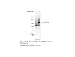
- Western Blot: HIF-1 alpha Antibody [NB100-654] - Panc0813 human pancreatic cell line. Image supplied by customer using Lot C-2.

- Submitted by
- Novus Biologicals (provider)
- Main image

- Experimental details
- Simple Western: HIF-1 alpha Antibody [NB100-654] - Simple Western lane view shows a specific band for HIF-1 alpha in 0.5 mg/ml of Hypoxic HeLa lysate. This experiment was performed under reducing conditions using the 12-230 kDa separation system.

- Submitted by
- Novus Biologicals (provider)
- Main image
- Experimental details
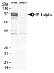
- Western Blot: HIF-1 alpha Antibody [NB100-654] - Detection of HIF-1 alpha in cobalt chloride treated/untreated COS-7 nuclear extracts using NB100-654.

- Submitted by
- Novus Biologicals (provider)
- Main image
- Experimental details
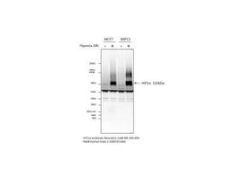
- Western Blot: HIF-1 alpha Antibody [NB100-654] - HIF1-alpha analysis using NB100-654. Image submitted from verified customer review.

- Submitted by
- Novus Biologicals (provider)
- Main image
- Experimental details
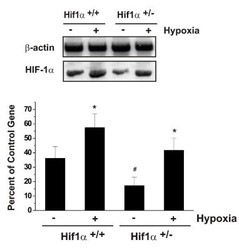
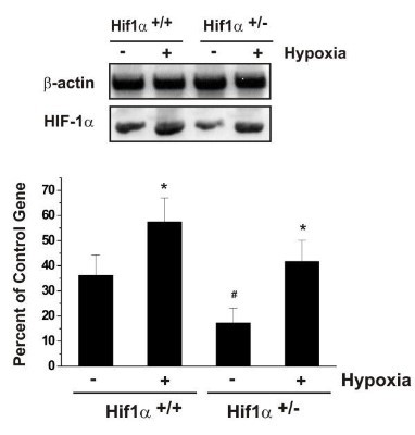
- Western Blot: HIF-1 alpha Antibody [NB100-654] - Effects of in vitro hypoxia on the expression of mouse HIF-1 alpha in HIF-1 alpha+/- and HIF-1 alpha+/+ astrocytes. Confluent astrocyte monolayers of both cell types were exposed to a 6 h in vitro hypoxia. HIF-1 alpha mRNA expression was determined by RT-PCR as described in Materials and Methods. Each bar represents the mean +/- SD of relative density/volumes of the bands on film negatives from at least three experiments. Asterisks and number sign indicate significant difference (p < 0.01; one-way ANOVA, followed by multiple comparisons among means). Image collected and cropped by CiteAb from the following publication (http://jneuroinflammation.biomedcentral.com/articles/10.1186/1742-2094-4-12), licensed under a CC-BY licence.
- Submitted by
- Novus Biologicals (provider)
- Main image

- Experimental details
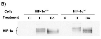
- Western Blot: HIF-1 alpha Antibody [NB100-654] - Effects of CoCl2 treatment on HIF-1 alpha expression in mouse astrocytes. Western blots using nuclear proteins show that both hypoxia (H) and CoCl2 (Co) up-regulated HIF-1 alpha protein in HIF-1 alpha+/+ and HIF-1 alpha+/- cells. There was no HIF-1 alpha protein detected in control cells. Image collected and cropped by CiteAb from the following publication (http://jneuroinflammation.biomedcentral.com/articles/10.1186/1742-2094-4-12), licensed under a CC-BY licence.
Supportive validation
- Submitted by
- Novus Biologicals (provider)
- Main image
- Experimental details
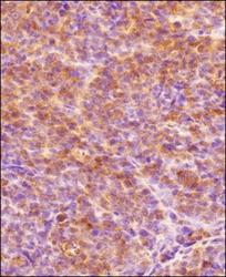
- Immunohistochemistry-Paraffin: HIF-1 alpha Antibody [NB100-654] - IHC analysis of a formalin-fixed paraffin-embedded tissue section of human endometrium carcinoma AN3CA cell line based xenograft using rabbit polyclonal HIF-1 alpha antibody NB100-654 at 1:300 dilution. The signal was developed using HRP-labelled secondary antibody and DAB reagent, and the section was further counterstained using hematoxylin. The tested section depicted mainly a diffused cytoplasmic staining but there were some cells which showed nuclear signal also (representing hypoxic cells).

Supportive validation
- Submitted by
- Novus Biologicals (provider)
- Main image
- Experimental details
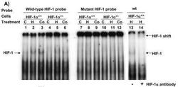
- Gel Super Shift Assays: HIF-1 alpha Antibody [NB100-654] - Mouse HIF-1 alpha+/+ and HIF-1 alpha+/- cells were exposed to hypoxia or 125 uM CoCl2 for 6 h, nuclear extracts were then prepared and EMSA carried out. HIF-1 isolated from hypoxia or CoCl2-treated cells bound to the wild-type probe (lanes #1-6) but not to the mutant probe (lanes #7-12). HIF-1/DNA complex was detected in hypoxia (H)-or CoCl2 (Co)-treated cells (lanes #2, 3, 5, 6) but not in control (C) cells (lanes #1, 4). More complex (darker band) was seen in hypoxia-or CoCl2-treated HIF-1 alpha+/+ cells (lanes #2, 3) than that in hypoxia-or CoCl2-treated HIF-1 alpha+/- cells (lanes #5, 6). Supershift assay showed that the HIF-1/DNA complex was shifted up in the presence of wild-type (wt) oligo probe and 4 ug HIF-1 alpha antiboby (lanes #13 & 14). Image collected and cropped by CiteAb from the following publication (http://jneuroinflammation.biomedcentral.com/articles/10.1186/1742-2094-4-12), licensed under a CC-BY licence.
