Explore
 Validate
 Learn
Learn Western blot
Antibody data
- Antibody Data
- Antigen structure
- References [0]
- Comments [0]
- Validations
- Western blot [2]
- Immunocytochemistry [1]
- Chromatin Immunoprecipitation [1]
- Other assay [1]
Submit
Validation data
Reference
Comment
Report error
- Product number
- 710059 - Provider product page

- Provider
- Invitrogen Antibodies
- Product name
- HIF1A Recombinant Polyclonal Antibody (16HCLC)
- Antibody type
- Polyclonal
- Antigen
- Other
- Description
- This antibody is predicted to react with mouse based on sequence homology.
- Antibody clone number
- 16HCLC
- Concentration
- 0.5 mg/mL
No comments: Submit comment
Supportive validation
- Submitted by
- Invitrogen Antibodies (provider)
- Main image
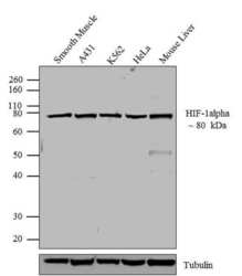
- Experimental details
- Western blot analysis of HIF1A was performed by loading 20 µg of A431 (lane 1), K562 (lane 2), HeLa (lane 3) and mouse liver (lane 4) cell lysates using Novex®NuPAGE®4-12 % Bis-Tris gel (Product # NP0321BOX), XCell SureLock Electrophoresis System (Product # EI0002), Novex® Sharp Pre-Stained Protein Standard (Product # LC5800), and iBlot® Dry Blotting System (Product # IB21001). Proteins were transferred to a nitrocellulose membrane and blocked with 5 % skim milk for 1 hour at room temperature. HIF1A was detected at ~80 kDa using HIF1A Recombinant Rabbit Polyclonal Antibody (Product # 710059) at 0.5-1 µg/mL in 2.5 % skim milk at 4°C overnight on a rocking platform. Goat anti-Rabbit IgG-HRP Secondary Antibody (Product # G-21234) at 1:5000 dilution was used and chemiluminescent detection was performed using Pierce™ ECL Western blotting Substrate (Product # 32106).

- Submitted by
- Invitrogen Antibodies (provider)
- Main image
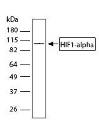
- Experimental details
- Western blot analysis of HIF-1 alpha in whole cell extract from HEK293 cells (30 µg per lane) using a HIF-1 alpha Recombinant Rabbit Polyclonal Antibody (Product # 710059) at a dilution of 1 µg/mL. Detection was performed using an HRP-conjugated Goat anti-Rabbit secondary antibody at a dilution of 1:5000 followed by chemiluminescence (ECL). Results show a band at ~93 kDa.
Supportive validation
- Submitted by
- Invitrogen Antibodies (provider)
- Main image
- Experimental details
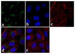
- Immunofluorescent analysis of HIF1-alpha was done on 70% confluent log phase U-2 OS cells. The cells were fixed with 4% paraformaldehyde for 15 minutes, and blocked with 5% BSA for 1 hour at room temperature. The cells were labeled with HIF1-alpha Recombinant Rabbit Polyclonal Antibody (Product # 710059) at 1 µg/mL in 1% BSA and incubated for 3 hours at room temperature and then labeled with Alexa Fluor 488 Goat anti-Rabbit IgG Secondary Antibody (Product # A-11008) at a dilution of 1:400 for 30 minutes at room temperature (Panel a: green). Nuclei (Panel b: blue) were stained with SlowFade Gold Antifade Mountant with DAPI (Product # S36938). F-actin (Panel c: red) was stained with Alexa Fluor 594 Phalloidin (Product # A12381). Panel d is a merged image showing cytoplasmic localization and panel e is a no primary antibody control. The images were captured at 20X magnification.

Supportive validation
- Submitted by
- Invitrogen Antibodies (provider)
- Main image
- Experimental details
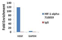
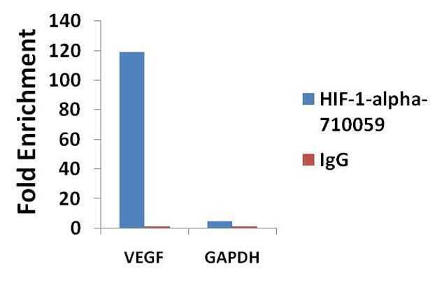
- ChIP- qPCR analysis of HIF1-alpha was performed with 3 µg/mL of the HIF1-alpha Recombinant Rabbit Polyclonal Antibody (Product # 710059) on sheared chromatin from 2 million HeLa cells serum starved overnight and serum released for 1h using the MAGnify Chromatin Immunoprecipitation System (Product # 49-2024). Normal rabbit IgG (3 µg/mL) was used as a negative IP control. The purified DNA from each ChIP sample was analyzed by StepOnePlus Real-Time PCR System (Product # 4376600) with primers for the promoter of active VEGF gene, used as positive control targets, and the GAPDH gene, used as negative control target. Data is presented as fold enrichment of the antibody signal versus the negative control IgG using the comparative CT method.
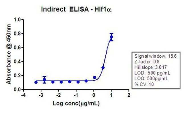
Supportive validation
- Submitted by
- Invitrogen Antibodies (provider)
- Main image

- Experimental details
- Indirect ELISA analysis of HIF-1 alpha in HEK293 cell lysate (300 ng/well) using a HIF-1 alpha Recombinant Rabbit Polyclonal Antibody (Product # 710059) and TMB (Product # SB01) as a substrate.