Explore
 Validate
 Learn
Learn14-9100-80
antibody from Invitrogen Antibodies
Targeting: HIF1A
bHLHe78, HIF-1alpha, HIF1, MOP1, PASD8
 Immunohistochemistry
Antibody data
- Antibody Data
- Antigen structure
- References [4]
- Comments [0]
- Validations
- Immunohistochemistry [1]
- Other assay [1]
Submit
Validation data
Reference
Comment
Report error
- Product number
- 14-9100-80 - Provider product page

- Provider
- Invitrogen Antibodies
- Product name
- HIF-1 alpha Monoclonal Antibody (ESEE122), eBioscience™
- Antibody type
- Monoclonal
- Antigen
- Other
- Description
- Description: This ESEE122 monoclonal antibody reacts with hypoxia inducible factor-1 alpha. The hypoxia inducible factor (HIF-1) is a transcription factor composed of an inducible alpha form and the constitutively expressed beta form, also called the aryl hydrocarbon receptor nuclear translocator (ARNT). Both alpha and beta subunits of HIF-1 contain basic helix-loop-helix motifs and function to activate transcription of genes in response to reduced oxygen levels. Under normoxic conditions, HIF-1 alpha is rapidly degraded, while HIF-1 alpha expression is induced and degradation inhibited under hypoxic conditions. HIF-1 alpha can be found in the cytoplasm and/or in the nucleus under normoxic conditions, but translocates to the nucleus under hypoxic conditions. HIF-1 is upregulated in several cancer types and functions to control genes involved in angiogenesis, cell survival and T cell development. Recent studies have shown the importance of HIF-1 alpha in balancing Th17 and Treg development. This ESEE122 antibody has also been reported to cross-react with bovine, mouse, and rat hypoxia inducible factor-1 alpha. Applications Reported: This ESEE122 antibody has been reported for use in immunohistochemical staining of formalin-fixed paraffin embedded tissue sections, and immunocytochemistry. Applications Tested: This ESEE122 antibody has been tested by immunohistochemistry on formalin-fixed paraffin embedded (FFPE) human placenta at less than or equal to 10 µg/mL using low pH antigen retrieval buffer. This ESEE122 antibody has been tested by immunocytochemistry on fixed and permeabilized Hela cells at less than or equal to 10 µg/mL. It is recommended that the antibody be titrated for optimal performance in the assay of interest. Purity: Greater than 90%, as determined by SDS-PAGE. Aggregation: Less than 10%, as determined by HPLC. Filtration: 0.2 µm post-manufacturing filtered.
- Reactivity
- Human, Mouse, Rat, Bovine
- Host
- Mouse
- Isotype
- IgG
- Antibody clone number
- ESEE122
- Vial size
- 25 µg
- Concentration
- 0.5 mg/mL
- Storage
- 4° C
Submitted references Gelatin hydrogel nonwoven fabrics of a cell culture scaffold to formulate 3-dimensional cell constructs.
Control of T(H)17/T(reg) balance by hypoxia-inducible factor 1.
Induction of the hypoxia-inducible factor system by low levels of heat shock protein 90 inhibitors.
Hypoxia-inducible factor induction by tumour necrosis factor in normoxic cells requires receptor-interacting protein-dependent nuclear factor kappa B activation.
Saotome T, Shimada N, Matsuno K, Nakamura K, Tabata Y
Regenerative therapy 2021 Dec;18:418-429
Regenerative therapy 2021 Dec;18:418-429
Control of T(H)17/T(reg) balance by hypoxia-inducible factor 1.
Dang EV, Barbi J, Yang HY, Jinasena D, Yu H, Zheng Y, Bordman Z, Fu J, Kim Y, Yen HR, Luo W, Zeller K, Shimoda L, Topalian SL, Semenza GL, Dang CV, Pardoll DM, Pan F
Cell 2011 Sep 2;146(5):772-84
Cell 2011 Sep 2;146(5):772-84
Induction of the hypoxia-inducible factor system by low levels of heat shock protein 90 inhibitors.
Ibrahim NO, Hahn T, Franke C, Stiehl DP, Wirthner R, Wenger RH, Katschinski DM
Cancer research 2005 Dec 1;65(23):11094-100
Cancer research 2005 Dec 1;65(23):11094-100
Hypoxia-inducible factor induction by tumour necrosis factor in normoxic cells requires receptor-interacting protein-dependent nuclear factor kappa B activation.
Jung Y, Isaacs JS, Lee S, Trepel J, Liu ZG, Neckers L
The Biochemical journal 2003 Mar 15;370(Pt 3):1011-7
The Biochemical journal 2003 Mar 15;370(Pt 3):1011-7
No comments: Submit comment
Supportive validation
- Submitted by
- Invitrogen Antibodies (provider)
- Main image
- Experimental details
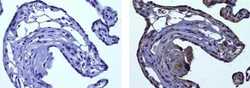
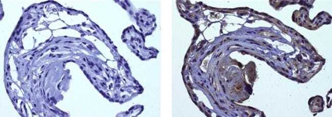
- Immunohistochemistry on formalin-fixed paraffin embedded human placenta, using 10 µg/mL Mouse IgG1 Isotype Control (left) or 10 µg/mL Anti-HIF-1 alpha Purified (right) followed by biotinylated Anti-Mouse IgG, and DAB visualization.Nuclei are counterstained with hematoxylin.
Supportive validation
- Submitted by
- Invitrogen Antibodies (provider)
- Main image
- Experimental details
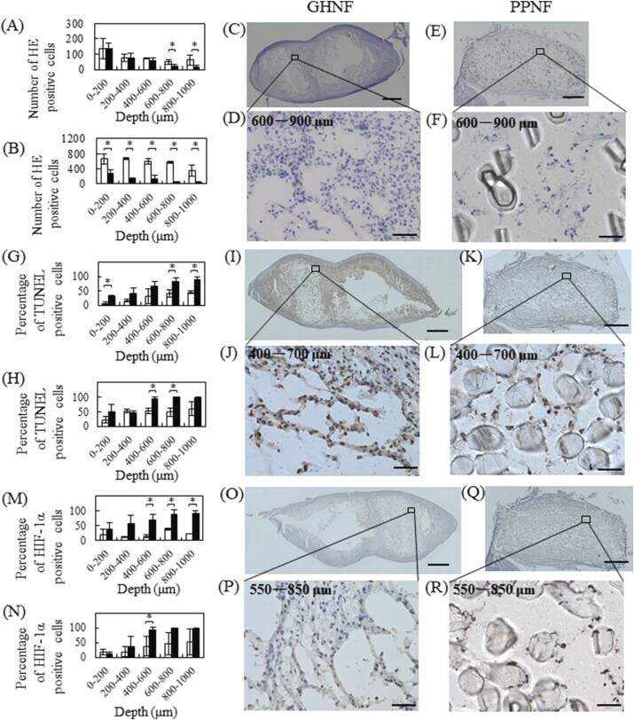
- Fig. 4 Distribution of HE-, TUNEL-, and HIF-1alpha-positive cells in GHNF and PPNF 14 and 35 days after stirring culture. Depth profiles of HE-positive cells in GHNF (#) and PPNF (#) 14 (A) and 35 days (B) after stirring culture. The cross-sectional images of GHNF (C, E) and PPNF (D, F) 35 days after stirring culture. HE images at 600-900 mum depth from the surface of GHNF (D) and PPNF (F). Depth profiles of TUNEL-positive cells in GHNF (#) and PPNF (#)14 (G) and 35 days after stirring culture (H). The cross-sectional images of GHNF (I, J) and PPNF (K, L) 35 days after stirring culture. TUNEL staining images at 400-700 mum depth from the surface of GHNF (J) and PPNF (L). Depth profiles of HIF-1alpha-positive cells in GHNF (#) and PPNF (#)14 (M) and 35 days after stirring culture (N). The cross-sectional images of GHNF (O, P) and PPNF (Q, R) 35 days after stirring culture. HIF-1alpha staining images at 550-850 mum depth from the surface of GHNF (P) and PPNF (R). The scale bar indicates 1 mm (C, E, I, K, O, Q) and 50 mum (D, F, J, L, P, R). *, p < 0.05; significant difference between the two groups. Fig. 4