Explore
 Validate
 Learn
LearnPA1-16627
antibody from Invitrogen Antibodies
Targeting: HIF1A
bHLHe78, HIF-1alpha, HIF1, MOP1, PASD8
 Western blot
 Immunocytochemistry
 Gel shift
Antibody data
- Antibody Data
- Antigen structure
- References [0]
- Comments [0]
- Validations
- Immunocytochemistry [2]
- Immunohistochemistry [1]
Submit
Validation data
Reference
Comment
Report error
- Product number
- PA1-16627 - Provider product page

- Provider
- Invitrogen Antibodies
- Product name
- HIF1A Polyclonal Antibody
- Antibody type
- Polyclonal
- Antigen
- Other
- Description
- This antibody has 93% sequence homology with bovine and 90% sequence homology with mouse. Suggested positive control: Cos-7 Hypoxia induced nuclear extract, COS-7 nuclear extracts (treated).
- Reactivity
- Human, Mouse, Rat, Bovine, Porcine
- Host
- Rabbit
- Isotype
- IgG
- Vial size
- 100 μL
- Concentration
- 1 mg/mL
- Storage
- -20°C, Avoid Freeze/Thaw Cycles
No comments: Submit comment
Supportive validation
- Submitted by
- Invitrogen Antibodies (provider)
- Main image
- Experimental details
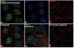
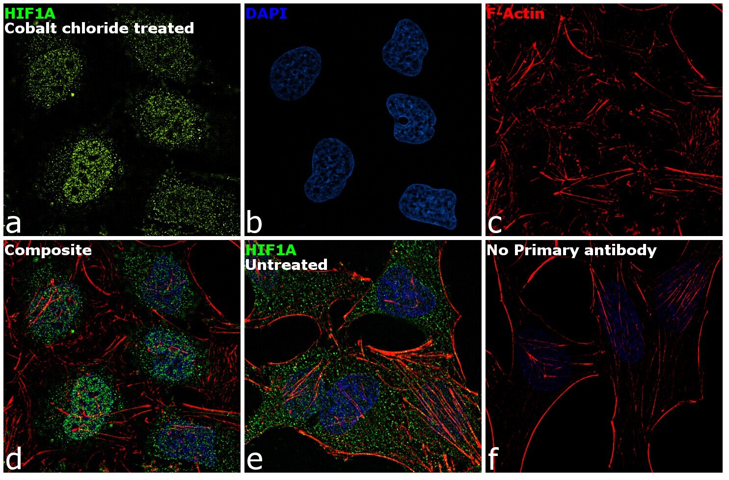
- Immunofluorescence analysis of HIF1A was performed using 70% confluent log phase HeLa cells treated with 150µM of CoCl2 for 48 hours. The cells were fixed with 4% paraformaldehyde for 10 minutes, permeabilized with 0.1% Triton™ X-100 for 15 minutes, and blocked with 1% BSA for 1 hour at room temperature. The cells were labeled with HIF1A Rabbit Polyclonal Antibody(Product # PA1-16627) at 5 µg/mL in 0.1% BSA, incubated at 4 degree Celsius overnight and then labeled with Goat anti-Rabbit IgG (H+L) Superclonal™ Secondary Antibody, Alexa Fluor® 488 conjugate (Product # A27034) at a dilution of 1:2000 for 45 minutes at room temperature (Panel a: green). Nuclei (Panel b: blue) were stained with SlowFade® Gold Antifade Mountant with DAPI (Product # S36938). F-actin (Panel c: red) was stained with Rhodamine Phalloidin (Product # R415, 1:300). Panel d represents the merged image showing nuclear localization. Panel e shows untreated cells with cytoplasmic signal. Panel f represents control cells with no primary antibody to assess background. The images were captured at 60X magnification.
- Submitted by
- Invitrogen Antibodies (provider)
- Main image

- Experimental details
- Immunofluorescence analysis of HIF1A was performed using 70% confluent log phase HeLa cells treated with 150µM of CoCl2 for 48 hours. The cells were fixed with 4% paraformaldehyde for 10 minutes, permeabilized with 0.1% Triton™ X-100 for 15 minutes, and blocked with 1% BSA for 1 hour at room temperature. The cells were labeled with HIF1A Rabbit Polyclonal Antibody(Product # PA1-16627) at 5 µg/mL in 0.1% BSA, incubated at 4 degree Celsius overnight and then labeled with Goat anti-Rabbit IgG (Heavy Chain) Superclonal™ Secondary Antibody, Alexa Fluor® 488 conjugate (Product # A27034) at a dilution of 1:2000 for 45 minutes at room temperature (Panel a: green). Nuclei (Panel b: blue) were stained with SlowFade® Gold Antifade Mountant with DAPI (Product # S36938). F-actin (Panel c: red) was stained with Rhodamine Phalloidin (Product # R415, 1:300). Panel d represents the merged image showing nuclear localization. Panel e shows untreated cells with cytoplasmic signal. Panel f represents control cells with no primary antibody to assess background. The images were captured at 60X magnification.

Supportive validation
- Submitted by
- Invitrogen Antibodies (provider)
- Main image
- Experimental details
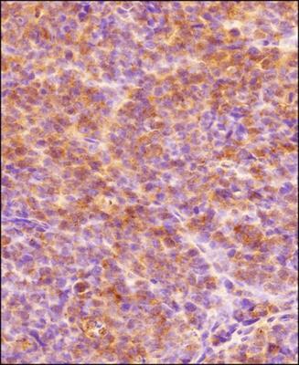
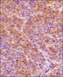
- Immunohistochemical analysis of HIF1A in formalin-fixed paraffin-embedded tissue section of human endometrium carcinoma AN3CA cell line based xenograft. Samples were incubated in HIF1A polyclonal antibody (Product # PA1-16627) using a dilution of 1:300. The signal was developed using HRP-labelled secondary antibody and DAB reagent, and the section was further counterstained using hematoxylin. The tested section depicted mainly a diffused cytoplasmic staining but there were some cells which showed nuclear signal also (representing hypoxic cells).