Explore
 Validate
 Learn
Learn Western blot
Antibody data
- Antibody Data
- Antigen structure
- References [4]
- Comments [0]
- Validations
- Western blot [1]
- Immunohistochemistry [1]
Submit
Validation data
Reference
Comment
Report error
- Product number
- AF5267 - Provider product page

- Provider
- Novus Biologicals
- Product name
- Sheep Polyclonal Notch-1 Antibody
- Antibody type
- Polyclonal
- Description
- Immunogen affinity purified. Detects mouse Notch-1 in direct ELISAs and Western blots. In direct ELISA, approximately 10% cross-reactivity with recombinant human Notch-1 and recombinant rat Notch-1 is observed and less than 1% cross-reactivity with recombinant mouse (rm) Notch-2 and rmNotch-3 is observed.
- Reactivity
- Mouse
- Host
- Sheep
- Isotype
- IgG
- Vial size
- 100 ug
- Concentration
- LYOPH
- Storage
- Use a manual defrost freezer and avoid repeated freeze-thaw cycles. 12 months from date of receipt, -20 to -70 degreesC as supplied. 1 month, 2 to 8 degreesC under sterile conditions after reconstitution. 6 months, -20 to -70 degreesC under sterile conditions after reconstitution.
Submitted references Notch signalling defines dorsal root ganglia neuroglial fate choice during early neural crest cell migration.
Two novel protein O-glucosyltransferases that modify sites distinct from POGLUT1 and affect Notch trafficking and signaling.
Uncontrolled angiogenic precursor expansion causes coronary artery anomalies in mice lacking Pofut1.
Antibodies that detect O-linked β-D-N-acetylglucosamine on the extracellular domain of cell surface glycoproteins.
Wiszniak S, Schwarz Q
BMC neuroscience 2019 Apr 29;20(1):21
BMC neuroscience 2019 Apr 29;20(1):21
Two novel protein O-glucosyltransferases that modify sites distinct from POGLUT1 and affect Notch trafficking and signaling.
Takeuchi H, Schneider M, Williamson DB, Ito A, Takeuchi M, Handford PA, Haltiwanger RS
Proceedings of the National Academy of Sciences of the United States of America 2018 Sep 4;115(36):E8395-E8402
Proceedings of the National Academy of Sciences of the United States of America 2018 Sep 4;115(36):E8395-E8402
Uncontrolled angiogenic precursor expansion causes coronary artery anomalies in mice lacking Pofut1.
Wang Y, Wu B, Lu P, Zhang D, Wu B, Varshney S, Del Monte-Nieto G, Zhuang Z, Charafeddine R, Kramer AH, Sibinga NE, Frangogiannis NG, Kitsis RN, Adams RH, Alitalo K, Sharp DJ, Harvey RP, Stanley P, Zhou B
Nature communications 2017 Sep 18;8(1):578
Nature communications 2017 Sep 18;8(1):578
Antibodies that detect O-linked β-D-N-acetylglucosamine on the extracellular domain of cell surface glycoproteins.
Tashima Y, Stanley P
The Journal of biological chemistry 2014 Apr 18;289(16):11132-11142
The Journal of biological chemistry 2014 Apr 18;289(16):11132-11142
No comments: Submit comment
Supportive validation
- Submitted by
- Novus Biologicals (provider)
- Main image

- Experimental details
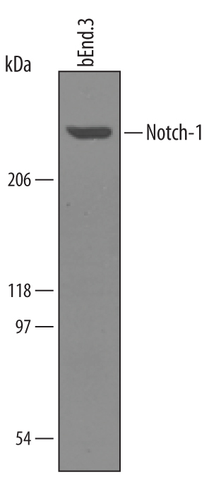
- Detection of Mouse Notch-1 by Western Blot. Western blot shows lysates of bEnd.3 mouse endothelioma cell line. PVDF membrane was probed with 1 µg/mL of Mouse Notch-1 Antigen Affinity-purified Polyclonal Antibody (Catalog # AF5267) followed by HRP-conjugated Anti-Sheep IgG Secondary Antibody (Catalog # HAF016). A specific band was detected for Notch-1 at approximately 300 kDa (as indicated). This experiment was conducted under reducing conditions and using Immunoblot Buffer Group 8.
Supportive validation
- Submitted by
- Novus Biologicals (provider)
- Main image
- Experimental details
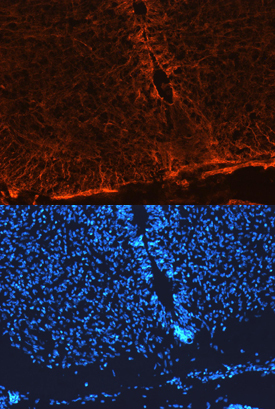
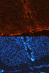
- Notch-1 in Mouse Neural Tube. Notch-1 was detected in immersion fixed frozen sections of mouse neural tube (E13.5) using Mouse Notch-1 Antigen Affinity-purified Polyclonal Antibody (Catalog # AF5267) at 10 µg/mL overnight at 4 °C. Tissue was stained using the NorthernLights™ 557-conjugated Anti-Sheep IgG Secondary Antibody (red, upper panel; Catalog # NL010) and counterstained with DAPI (blue, lower panel). View our protocol for Fluorescent IHC Staining of Frozen Tissue Sections.