Explore
 Validate
 Learn
Learn Western blot
 Immunocytochemistry
Antibody data
- Antibody Data
- Antigen structure
- References [6]
- Comments [0]
- Validations
- Immunocytochemistry [2]
- Immunohistochemistry [1]
Submit
Validation data
Reference
Comment
Report error
- Product number
- AF1057 - Provider product page

- Provider
- R&D Systems
- Product name
- Mouse/Rat Notch-1 Antibody
- Antibody type
- Polyclonal
- Description
- Antigen Affinity-purified. Detects rat Notch-1 in direct ELISAs and Western blots. In direct ELISAs and Western blots, less than 5% cross-reactivity with recombinant rat Notch-2 and recombinant mouse Notch-3 is observed.
- Reactivity
- Mouse, Rat
- Host
- Goat
- Conjugate
- Unconjugated
- Antigen sequence
Q07008- Isotype
- IgG
- Vial size
- 100 ug
- Concentration
- LYOPH
- Storage
- Use a manual defrost freezer and avoid repeated freeze-thaw cycles. 12 months from date of receipt, -20 to -70 °C as supplied. 1 month, 2 to 8 °C under sterile conditions after reconstitution. 6 months, -20 to -70 °C under sterile conditions after reconstitution.
Submitted references Muscle Satellite Cell Cross-Talk with a Vascular Niche Maintains Quiescence via VEGF and Notch Signaling.
Notch signaling pathway is a potential therapeutic target for extracranial vascular malformations.
Cell-Cell Contact Area Affects Notch Signaling and Notch-Dependent Patterning.
Macrophage-dependent tumor cell transendothelial migration is mediated by Notch1/MenaINV-initiated invadopodium formation.
Human stem/progenitor cells from bone marrow enhance glial differentiation of rat neural stem cells: a role for transforming growth factor β and Notch signaling.
Distinct stages of myelination regulated by gamma-secretase and astrocytes in a rapidly myelinating CNS coculture system.
Verma M, Asakura Y, Murakonda BSR, Pengo T, Latroche C, Chazaud B, McLoon LK, Asakura A
Cell stem cell 2018 Oct 4;23(4):530-543.e9
Cell stem cell 2018 Oct 4;23(4):530-543.e9
Notch signaling pathway is a potential therapeutic target for extracranial vascular malformations.
Davis RB, Pahl K, Datto NC, Smith SV, Shawber C, Caron KM, Blatt J
Scientific reports 2018 Dec 20;8(1):17987
Scientific reports 2018 Dec 20;8(1):17987
Cell-Cell Contact Area Affects Notch Signaling and Notch-Dependent Patterning.
Shaya O, Binshtok U, Hersch M, Rivkin D, Weinreb S, Amir-Zilberstein L, Khamaisi B, Oppenheim O, Desai RA, Goodyear RJ, Richardson GP, Chen CS, Sprinzak D
Developmental cell 2017 Mar 13;40(5):505-511.e6
Developmental cell 2017 Mar 13;40(5):505-511.e6
Macrophage-dependent tumor cell transendothelial migration is mediated by Notch1/MenaINV-initiated invadopodium formation.
Pignatelli J, Bravo-Cordero JJ, Roh-Johnson M, Gandhi SJ, Wang Y, Chen X, Eddy RJ, Xue A, Singer RH, Hodgson L, Oktay MH, Condeelis JS
Scientific reports 2016 Nov 30;6:37874
Scientific reports 2016 Nov 30;6:37874
Human stem/progenitor cells from bone marrow enhance glial differentiation of rat neural stem cells: a role for transforming growth factor β and Notch signaling.
Robinson AP, Foraker JE, Ylostalo J, Prockop DJ
Stem cells and development 2011 Feb;20(2):289-300
Stem cells and development 2011 Feb;20(2):289-300
Distinct stages of myelination regulated by gamma-secretase and astrocytes in a rapidly myelinating CNS coculture system.
Watkins TA, Emery B, Mulinyawe S, Barres BA
Neuron 2008 Nov 26;60(4):555-69
Neuron 2008 Nov 26;60(4):555-69
No comments: Submit comment
Supportive validation
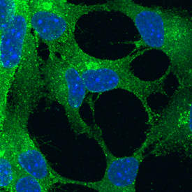
- Submitted by
- R&D Systems (provider)
- Main image

- Experimental details
- Notch-1 in Mouse Cortical Stem Cells. Notch-1 was detected in immersion fixed undifferentiated mouse cortical stem cells using Goat Anti-Mouse/Rat Notch-1 Antigen Affinity-purified Polyclonal Antibody (Catalog # AF1057) at 10 µg/mL for 3 hours at room temperature. Cells were stained using the NorthernLights™ 493-conjugated Anti-Goat IgG Secondary Antibody (green; Catalog # NL003) and counterstained with DAPI (blue). Specific staining was localized to cell surfaces. View our protocol for Fluorescent ICC Staining of Stem Cells on Coverslips.
- Submitted by
- R&D Systems (provider)
- Main image
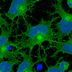
- Experimental details
- Notch-1 in Rat Cortical Stem Cells. Notch-1 was detected in immersion fixed undifferentiated rat cortical stem cells using Goat Anti-Mouse/Rat Notch-1 Antigen Affinity-purified Polyclonal Antibody (Catalog # AF1057) at 10 µg/mL for 3 hours at room temperature. Cells were stained using the NorthernLights™ 493-conjugated Anti-Goat IgG Secondary Antibody (green; Catalog # NL003) and counterstained with DAPI (blue). Specific staining was localized to cell surfaces. View our protocol for Fluorescent ICC Staining of Stem Cells on Coverslips.

Supportive validation
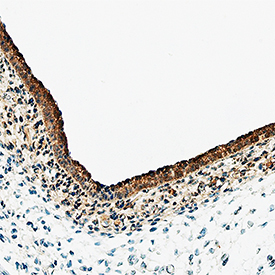
- Submitted by
- R&D Systems (provider)
- Main image

- Experimental details
- Notch-1 in Rat Embryo. Notch-1 was detected in immersion fixed paraffin-embedded sections of rat embryo (13 d.p.c.) using Goat Anti-Mouse/Rat Notch-1 Antigen Affinity-purified Polyclonal Antibody (Catalog # AF1057) at 10 µg/mL overnight at 4 °C. Tissue was stained using the Anti-Goat HRP-DAB Cell & Tissue Staining Kit (brown; Catalog # CTS008) and counterstained with hematoxylin (blue). Specific staining was localized to labeling is in epidermis. View our protocol for Chromogenic IHC Staining of Paraffin-embedded Tissue Sections.