 Explore
 Validate
 Learn
Learn Western blot
 Immunohistochemistry
 Other assay
Antibody data
- Antibody Data
- Antigen structure
- References [9]
- Comments [0]
- Validations
- Other assay [3]
Submit
Validation data
Reference
Comment
Report error
- Product number
- 14-5785-37 - Provider product page

- Provider
- Invitrogen Antibodies
- Product name
- NOTCH1 Monoclonal Antibody (mN1A), eBioscience™
- Antibody type
- Monoclonal
- Antigen
- Other
- Description
- Description: The Notch family of transmembrane receptors controls cell-fate decisions during the development of many organs in a wide variety of species. After binding its ligand, the Notch receptor is cleaved in its transmembrane domain, and the resulting intracellular domain dissociates from the membrane and translocates to the nucleus, where it is able to suppress the expression of lineage-specific genes by interacting with transcriptional repressors. The mN1A antibody reacts with the intracellular domain of mouse and human Notch1, but not with Notch2, 3, or 4. The mN1A antibody has a low affinity for the full-length (unprocessed or heterodimeric cell surface) forms of Notch1. In the mouse, Notch mRNA is expressed in mouse hematopoietic cells of the fetal liver and adult thymus and bone marrow. In the thymus, Notch1 protein is detected in CD4-CD8- (double-negative) and CD4-CD8+ (single-positive) thymocytes. Studies of Notch1-transgenic cells and Notch1-null mice indicate that the receptor is involved in the regulation of lymphopoiesis and myelopoiesis. Applications Reported: This mN1A antibody has been reported for use in intracellular staining followed by flow cytometric analysis, immunoblotting (WB), and immunohistochemical staining. (Fluorochrome conjugated mN1A is recommended for use in intracellular flow cytometry.). Applications Tested: This mN1A antibody has been tested by intracellular staining and flow cytometric analysis of mouse thymocytes. This can be used at less than or equal to 1 µg per test. A test is defined as the amount (µg) of antibody that will stain a cell sample in a final volume of 100 µL. Cell number should be determined empirically but can range from 10^5 to 10^8 cells/test. It is recommended that the antibody be carefully titrated for optimal performance in the assay of interest. Purity: Greater than 90%, as determined by SDS-PAGE. Aggregation: Less than 10%, as determined by HPLC. Filtration: 0.2 µm post-manufacturing filtered.
- Reactivity
- Human, Mouse
- Host
- Mouse
- Isotype
- IgG
- Antibody clone number
- mN1A
- Vial size
- 2 mg
- Concentration
- 0.5 mg/mL
- Storage
- 4°C
Submitted references The c-Myc Oncogene Maintains Corneal Epithelial Architecture at Homeostasis, Modulates p63 Expression, and Enhances Proliferation During Tissue Repair.
Genetic alterations of Keap1 confers chemotherapeutic resistance through functional activation of Nrf2 and Notch pathway in head and neck squamous cell carcinoma.
Whole Exome Sequencing reveals NOTCH1 mutations in anaplastic large cell lymphoma and points to Notch both as a key pathway and a potential therapeutic target.
FOXP3 expression is modulated by TGF‑β1/NOTCH1 pathway in human melanoma.
Therapeutic targeting of NOTCH signaling ameliorates immune-mediated bone marrow failure of aplastic anemia.
Novel protein transduction domain mimics as nonviral delivery vectors for siRNA targeting NOTCH1 in primary human T cells.
Dll4-Notch signaling in Flt3-independent dendritic cell development and autoimmunity in mice.
Notch signaling regulates mouse and human Th17 differentiation.
Pathways contributing to development of spontaneous mammary tumors in BALB/c-Trp53+/- mice.
Portal C, Wang Z, Scott DK, Wolosin JM, Iomini C
Investigative ophthalmology & visual science 2022 Feb 1;63(2):3
Investigative ophthalmology & visual science 2022 Feb 1;63(2):3
Genetic alterations of Keap1 confers chemotherapeutic resistance through functional activation of Nrf2 and Notch pathway in head and neck squamous cell carcinoma.
Islam SS, Qassem K, Islam S, Parag RR, Rahman MZ, Farhat WA, Yeger H, Aboussekhra A, Karakas B, Noman ASM
Cell death & disease 2022 Aug 9;13(8):696
Cell death & disease 2022 Aug 9;13(8):696
Whole Exome Sequencing reveals NOTCH1 mutations in anaplastic large cell lymphoma and points to Notch both as a key pathway and a potential therapeutic target.
Larose H, Prokoph N, Matthews JD, Schlederer M, Högler S, Alsulami AF, Ducray SP, Nuglozeh E, Fazaludeen FMS, Elmouna A, Ceccon M, Mologni L, Gambacorti-Passerini C, Hoefler G, Lobello C, Pospisilova S, Janikova A, Woessmann W, Damm-Welk C, Zimmermann M, Federova A, Malone A, Smith O, Wasik M, Inghirami G, Lamant L, Blundell TL, Klapper W, Merkel O, Burke AGA, Mian S, Ashankyty I, Kenner L, Turner SD
Haematologica 2021 Jun 1;106(6):1693-1704
Haematologica 2021 Jun 1;106(6):1693-1704
FOXP3 expression is modulated by TGF‑β1/NOTCH1 pathway in human melanoma.
Skarmoutsou E, Bevelacqua V, D' Amico F, Russo A, Spandidos DA, Scalisi A, Malaponte G, Guarneri C
International journal of molecular medicine 2018 Jul;42(1):392-404
International journal of molecular medicine 2018 Jul;42(1):392-404
Therapeutic targeting of NOTCH signaling ameliorates immune-mediated bone marrow failure of aplastic anemia.
Roderick JE, Gonzalez-Perez G, Kuksin CA, Dongre A, Roberts ER, Srinivasan J, Andrzejewski C Jr, Fauq AH, Golde TE, Miele L, Minter LM
The Journal of experimental medicine 2013 Jul 1;210(7):1311-29
The Journal of experimental medicine 2013 Jul 1;210(7):1311-29
Novel protein transduction domain mimics as nonviral delivery vectors for siRNA targeting NOTCH1 in primary human T cells.
Tezgel AÖ, Gonzalez-Perez G, Telfer JC, Osborne BA, Minter LM, Tew GN
Molecular therapy : the journal of the American Society of Gene Therapy 2013 Jan;21(1):201-9
Molecular therapy : the journal of the American Society of Gene Therapy 2013 Jan;21(1):201-9
Dll4-Notch signaling in Flt3-independent dendritic cell development and autoimmunity in mice.
Billiard F, Lobry C, Darrasse-Jèze G, Waite J, Liu X, Mouquet H, DaNave A, Tait M, Idoyaga J, Leboeuf M, Kyratsous CA, Burton J, Kalter J, Klinakis A, Zhang W, Thurston G, Merad M, Steinman RM, Murphy AJ, Yancopoulos GD, Aifantis I, Skokos D
The Journal of experimental medicine 2012 May 7;209(5):1011-28
The Journal of experimental medicine 2012 May 7;209(5):1011-28
Notch signaling regulates mouse and human Th17 differentiation.
Keerthivasan S, Suleiman R, Lawlor R, Roderick J, Bates T, Minter L, Anguita J, Juncadella I, Nickoloff BJ, Le Poole IC, Miele L, Osborne BA
Journal of immunology (Baltimore, Md. : 1950) 2011 Jul 15;187(2):692-701
Journal of immunology (Baltimore, Md. : 1950) 2011 Jul 15;187(2):692-701
Pathways contributing to development of spontaneous mammary tumors in BALB/c-Trp53+/- mice.
Yan H, Blackburn AC, McLary SC, Tao L, Roberts AL, Xavier EA, Dickinson ES, Seo JH, Arenas RB, Otis CN, Cao QJ, Lawlor RG, Osborne BA, Kittrell FS, Medina D, Jerry DJ
The American journal of pathology 2010 Mar;176(3):1421-32
The American journal of pathology 2010 Mar;176(3):1421-32
No comments: Submit comment
Supportive validation
- Submitted by
- Invitrogen Antibodies (provider)
- Main image
- Experimental details
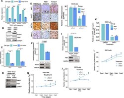
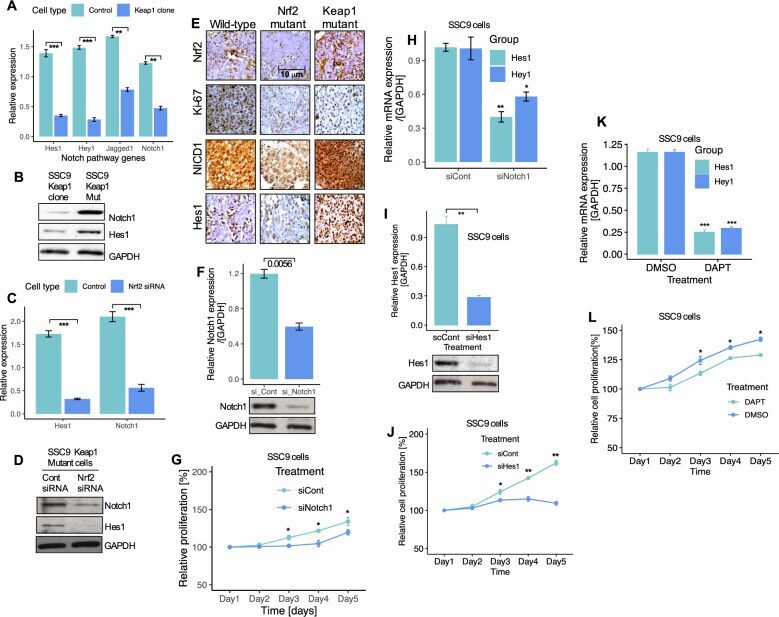
- Nrf2 regulates Notch signaling in HNSCC cells. A Expression of Notch1 and Notch target genes mRNA in control and Keap1- expressing SSC9 clone cells. B Expression of Notch1 and Hes1 proteins in Keap1 -mutant and Keap1- expressing SSC9 cells. C Notch1 and Hes1 mRNA and, D protein expression after Nrf2 knockdown in SSC9 cells. E Immunohistochemistry staining and expression of Nrf2 , Ki67, Notch1, and Hes1 in HNSCC clinical samples from wild-type, Nrf2 , and Keap1 mutant patients tumor tissues. F Notch1 expression in non-targeting control and Notch1 siRNA-treated SSC9 cells G Cell proliferation of SSC9 cells after knockdown of Notch1 by siRNA. H Relative mRNA expression of Hes1 and Hey1 after Notch1 knockdown in SSC9 cells. I Hes1 mRNA expression and, J Cell proliferation after knockdown of Hes1 siRNA in SSC9 cells. K Effects of Notch inhibitor DAPT and, L Assessment of cell growth after treating the cells with Notch inhibitor DAPT for 5-days. The mRNA expression levels were calculated and normalized relative to GAPDH. All experiments were run in triplicate and compared with the control group. Data presents as mean +- SEM (** P < 0.01, *** P < 0.001).
- Submitted by
- Invitrogen Antibodies (provider)
- Main image
- Experimental details
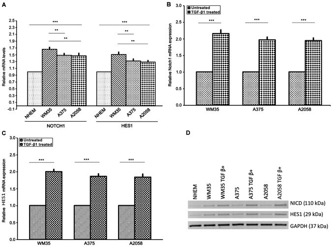
- Figure 4 Expression of NOTCH1 NICD and NOTCH-specific target gene HES1 in melanoma cell lines. (A) mRNA of NOTCH1 NICD and HES1 was measured by RT-qPCR in NHEM, WM35 and A375 and A2058 melanoma cells. Melanocytes served as the control. WM35 showed a higher level of NOTCH1 NICD mRNA and HES1 mRNA than A375 and A2058 cells. (B) Protein level of NOTCH1 NICD and HES1 was measured by western blot analysis in WM35, A375 and A2058 melanoma cell lines. All of the melanoma cell lines positively expressed NOTCH1 NICD and HES1. (B-D) Effect of TGFbeta-1 treatment on NOTCH1 NICD , HES1 mRNA and protein levels in melanoma cell lines. Treatment with rhTGF-beta1 (5 ng/ml) for 48 h induced a higher increase of NOTCH1 NICD and HES1 mRNA and their own protein levels in WM35, A375 and A2058 melanoma cells. As an internal control, GAPDH was used for normalization. Data are shown as mean +- SD of three independent experiments. The comparison of mRNA NOTCH-1 and HES1 expression in multiple groups was performed by ANOVA and Tukey's test. HES1, hairy and enhancer of split 1; RT-qPCR, reverse transcription-quantitative polymerase chain reaction; TGF-beta, transforming growth factor-beta; rh, recombinant human; GAPDH, glyceraldehyde 3-phosphate dehydrogenase. ** P
- Submitted by
- Invitrogen Antibodies (provider)
- Main image
- Experimental details
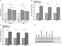
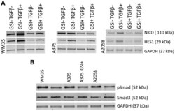
- Figure 5 GSI effect on NOTCH1 NICD , NOTCH-specific target gene HES1 expression and on TGF-beta/Smad signaling in melanoma cell lines. (A) Inhibition of NOTCH1 NICD and NOTCH-specific target gene HES1 is illustrated after 72 h of 20 mu M GSI treatment in WM35, A375 and A2058 cells. Western blot analysis showed that GSI suppressed NOTCH1 NICD and HES1 protein levels and downregulated TGFbeta-1- induced NOTCH1 NICD , HES1 protein levels in melanoma cell lines. (B) GSI treatment consistently decreased pSMAD3 levels in all melanoma cell lines. GAPDH served as loading control. GSI, gamma-secretase inhibitor; HES1, hairy and enhancer of split 1; TGF-beta, transforming growth factor-beta; pSMAD3, phosphorylated Smad3; GAPDH, glyceraldehyde 3-phosphate dehydrogenase.
