Explore
 Validate
 Learn
Learn Western blot
 Immunocytochemistry
Antibody data
- Antibody Data
- Antigen structure
- References [7]
- Comments [0]
- Validations
- Western blot [1]
- Immunohistochemistry [1]
- Flow cytometry [1]
Submit
Validation data
Reference
Comment
Report error
- Product number
- NB100-78486 - Provider product page

- Provider
- Novus Biologicals
- Proper citation
- Novus Cat#NB100-78486, RRID:AB_1085196
- Product name
- Mouse Monoclonal Notch-1 Antibody
- Antibody type
- Monoclonal
- Description
- Protein G purified. Does not cross-react with Notch 2, 3, or 4.
- Reactivity
- Human, Mouse, Rat
- Host
- Mouse
- Isotype
- IgG
- Vial size
- 0.1 ml
- Concentration
- 1.0 mg/ml
- Storage
- Store at 4C short term. Aliquot and store at -20C long term. Avoid freeze-thaw cycles.
Submitted references Osteoblast Hypoxia-Inducible Factor-1α Pathway Activation Restrains Osteoclastogenesis via the Interleukin-33-MicroRNA-34a-Notch1 Pathway.
Cyclic AMP Response Element Binding Protein Mediates Pathological Retinal Neovascularization via Modulating DLL4-NOTCH1 Signaling.
Aberrant expression of Notch1 interferes with the B-lymphoid phenotype of neoplastic B cells in classical Hodgkin lymphoma.
Stra13 regulates satellite cell activation by antagonizing Notch signaling.
Evidence for a physical interaction between presenilin and Notch.
Evidence for a physical interaction between presenilin and Notch.
A presenilin-1-dependent gamma-secretase-like protease mediates release of Notch intracellular domain.
Kang H, Yang K, Xiao L, Guo L, Guo C, Yan Y, Qi J, Wang F, Ryffel B, Li C, Deng L
Frontiers in immunology 2017;8:1312
Frontiers in immunology 2017;8:1312
Cyclic AMP Response Element Binding Protein Mediates Pathological Retinal Neovascularization via Modulating DLL4-NOTCH1 Signaling.
Singh NK, Kotla S, Kumar R, Rao GN
EBioMedicine 2015 Nov;2(11):1767-84
EBioMedicine 2015 Nov;2(11):1767-84
Aberrant expression of Notch1 interferes with the B-lymphoid phenotype of neoplastic B cells in classical Hodgkin lymphoma.
Jundt F, Acikgöz O, Kwon SH, Schwarzer R, Anagnostopoulos I, Wiesner B, Mathas S, Hummel M, Stein H, Reichardt HM, Dörken B
Leukemia 2008 Aug;22(8):1587-94
Leukemia 2008 Aug;22(8):1587-94
Stra13 regulates satellite cell activation by antagonizing Notch signaling.
Sun H, Li L, Vercherat C, Gulbagci NT, Acharjee S, Li J, Chung TK, Thin TH, Taneja R
The Journal of cell biology 2007 May 21;177(4):647-57
The Journal of cell biology 2007 May 21;177(4):647-57
Evidence for a physical interaction between presenilin and Notch.
Ray WJ, Yao M, Nowotny P, Mumm J, Zhang W, Wu JY, Kopan R, Goate AM
Proceedings of the National Academy of Sciences of the United States of America 1999 Mar 16;96(6):3263-8
Proceedings of the National Academy of Sciences of the United States of America 1999 Mar 16;96(6):3263-8
A presenilin-1-dependent gamma-secretase-like protease mediates release of Notch intracellular domain.
De Strooper B, Annaert W, Cupers P, Saftig P, Craessaerts K, Mumm JS, Schroeter EH, Schrijvers V, Wolfe MS, Ray WJ, Goate A, Kopan R
Nature 1999 Apr 8;398(6727):518-22
Nature 1999 Apr 8;398(6727):518-22
No comments: Submit comment
Supportive validation
- Submitted by
- Novus Biologicals (provider)
- Main image
- Experimental details
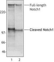
- Western Blot: Notch-1 Antibody (mN1A) [NB100-78486] - Cell extracts from Jurkat (Lane 1) or mouse thymocytes (Lane 2) were analyzed with monoclonal anti-NOTCH1 antibody. The mN1A antibody recognizes both mouse and human 270 kDa full-length NOTCH1 and 110-120 kDa cleaved NOTCH 1 (NICD).

Supportive validation
- Submitted by
- Novus Biologicals (provider)
- Main image
- Experimental details
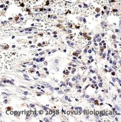
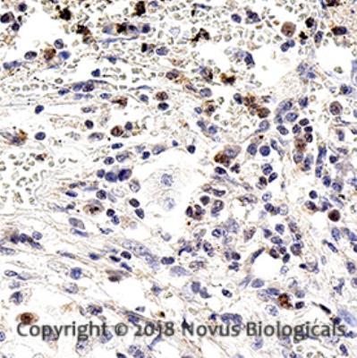
- Immunohistochemistry-Paraffin: Notch-1 Antibody (mN1A) [NB100-78486] - Analysis of FFPE human pancreatic cancer using 1:10 dilution of Notch-1 antibody on a Bond Rx autostainer (Leica Biosystems). The assay involved 20 minutes of heat induced antigen retrieval (HIER) using 10mM sodium citrate buffer (pH 6.0) and endogenous peroxidase quenching with peroxide block. The sections were incubated with primary antibody for 30 minutes and Bond Polymer Refine Detection (Leica Biosystems) with DAB was used for signal development followed by counterstaining with hematoxylin. Whole slide scanning and capturing of representative images (20X) was performed using Aperio AT2 (Leica Biosystems). Cytoplasmic staining in epithelial cells was observed. Staining was performed by Histowiz.
Supportive validation
- Submitted by
- Novus Biologicals (provider)
- Main image
- Experimental details
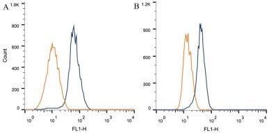
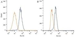
- Flow Cytometry: Notch-1 Antibody (mN1A) [NB100-78486] - Intracellular flow cytometric staining of 1 x 10^6 CHO (A) and MCF-7 (B) cells using Notch1 antibody (dark blue). Isotype control shown in orange. An antibody concentration of 1 ug/1x10^6 cells was used.