 Explore
 Validate
 Learn
Learn Western blot
 Immunocytochemistry
Antibody data
- Antibody Data
- Antigen structure
- References [1]
- Comments [0]
- Validations
- Immunocytochemistry [5]
- Immunohistochemistry [5]
- Flow cytometry [2]
- Other assay [4]
Submit
Validation data
Reference
Comment
Report error
- Product number
- PA5-85956 - Provider product page

- Provider
- Invitrogen Antibodies
- Product name
- BMP-2 Polyclonal Antibody
- Antibody type
- Polyclonal
- Antigen
- Synthetic peptide
- Reactivity
- Human, Mouse, Rat
- Host
- Rabbit
- Isotype
- IgG
- Vial size
- 100 μL
- Concentration
- 1 mg/mL
- Storage
- Store at 4°C short term. For long term storage, store at -20°C, avoiding freeze/thaw cycles.
Submitted references Femoral µCT Analysis, Mechanical Testing and Immunolocalization of Bone Proteins in β-Hydroxy β-Methylbutyrate (HMB) Supplemented Spiny Mouse in a Model of Pregnancy and Lactation-Associated Osteoporosis.
Tomaszewska E, Muszyński S, Donaldson J, Dobrowolski P, Chand DKP, Tomczyk-Warunek A, Hułas-Stasiak M, Puzio I, Lamorski K, Sławiński C, Jabłoński M, Blicharski T
Journal of clinical medicine 2021 Oct 20;10(21)
Journal of clinical medicine 2021 Oct 20;10(21)
No comments: Submit comment
Supportive validation
- Submitted by
- Invitrogen Antibodies (provider)
- Main image

- Experimental details
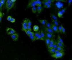
- Immunocytochemical analysis of BMP-2 in Hela cells using a BMP-2 Polyclonal antibody (Product # PA5-85956) as seen in green. Cells were fixed in paraformaldehyde, permeabilised with 0.25% Triton X100/PBS. DAPI was used to stain the cell nuclei (blue).

- Submitted by
- Invitrogen Antibodies (provider)
- Main image
- Experimental details
- Immunocytochemical analysis of BMP-2 in Hela cells using a BMP-2 Polyclonal antibody (Product # PA5-85956) as seen in green. Cells were fixed in paraformaldehyde, permeabilised with 0.25% Triton X100/PBS. DAPI was used to stain the cell nuclei (blue).

- Submitted by
- Invitrogen Antibodies (provider)
- Main image

- Experimental details
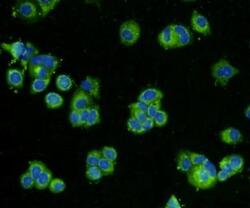
- Immunocytochemical analysis of BMP-2 in MCF-7 cells using a BMP-2 Polyclonal antibody (Product # PA5-85956) as seen in green. Cells were fixed in paraformaldehyde, permeabilised with 0.25% Triton X100/PBS. DAPI was used to stain the cell nuclei (blue).

- Submitted by
- Invitrogen Antibodies (provider)
- Main image

- Experimental details
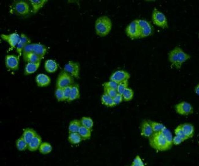
- Immunocytochemical analysis of BMP-2 in Hela cells using a BMP-2 Polyclonal antibody (Product # PA5-85956) as seen in green. Cells were fixed in paraformaldehyde, permeabilised with 0.25% Triton X100/PBS. DAPI was used to stain the cell nuclei (blue).

- Submitted by
- Invitrogen Antibodies (provider)
- Main image
- Experimental details
- Immunocytochemical analysis of BMP-2 in MCF-7 cells using a BMP-2 Polyclonal antibody (Product # PA5-85956) as seen in green. Cells were fixed in paraformaldehyde, permeabilised with 0.25% Triton X100/PBS. DAPI was used to stain the cell nuclei (blue).
Supportive validation
- Submitted by
- Invitrogen Antibodies (provider)
- Main image
- Experimental details
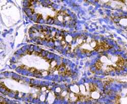
- Immunohistochemical analysis of BMP-2 of paraffin-embedded rat small intestine tissue using a BMP-2 Polyclonal antibody (Product #PA5-85956). Counter stained with hematoxylin.
- Submitted by
- Invitrogen Antibodies (provider)
- Main image

- Experimental details
- Immunohistochemical analysis of BMP-2 of paraffin-embedded Human breast cancer tissue using a BMP-2 Polyclonal antibody (Product #PA5-85956). Counter stained with hematoxylin.

- Submitted by
- Invitrogen Antibodies (provider)
- Main image
- Experimental details
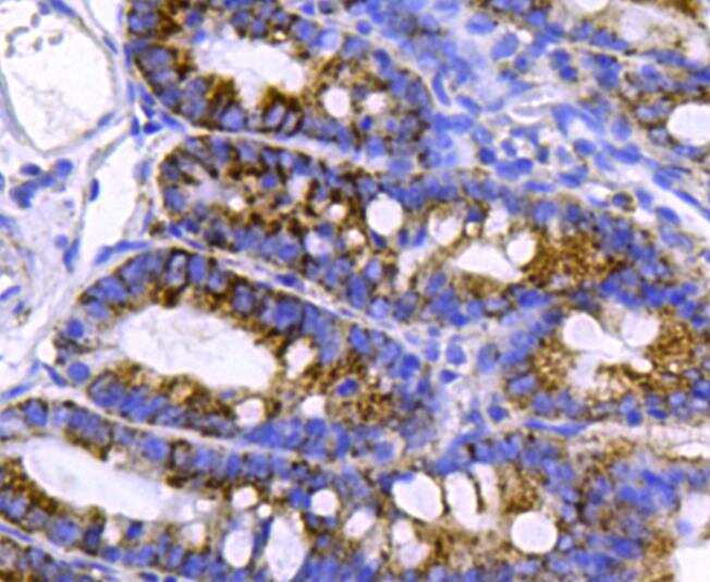
- Immunohistochemical analysis of BMP-2 of paraffin-embedded Mouse small intestine tissue using a BMP-2 Polyclonal antibody (Product #PA5-85956). Counter stained with hematoxylin.

- Submitted by
- Invitrogen Antibodies (provider)
- Main image

- Experimental details
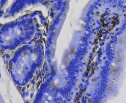
- Immunohistochemical analysis of BMP-2 of paraffin-embedded rat small intestine tissue using a BMP-2 Polyclonal antibody (Product #PA5-85956). Counter stained with hematoxylin.

- Submitted by
- Invitrogen Antibodies (provider)
- Main image

- Experimental details
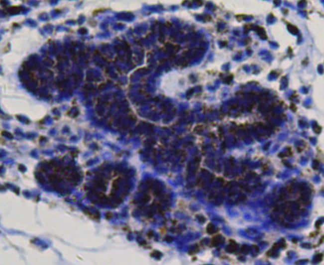
- Immunohistochemical analysis of BMP-2 of paraffin-embedded Human breast cancer tissue using a BMP-2 Polyclonal antibody (Product #PA5-85956). Counter stained with hematoxylin.
Supportive validation
- Submitted by
- Invitrogen Antibodies (provider)
- Main image
- Experimental details
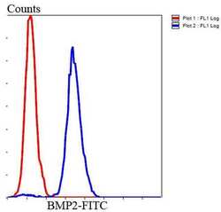
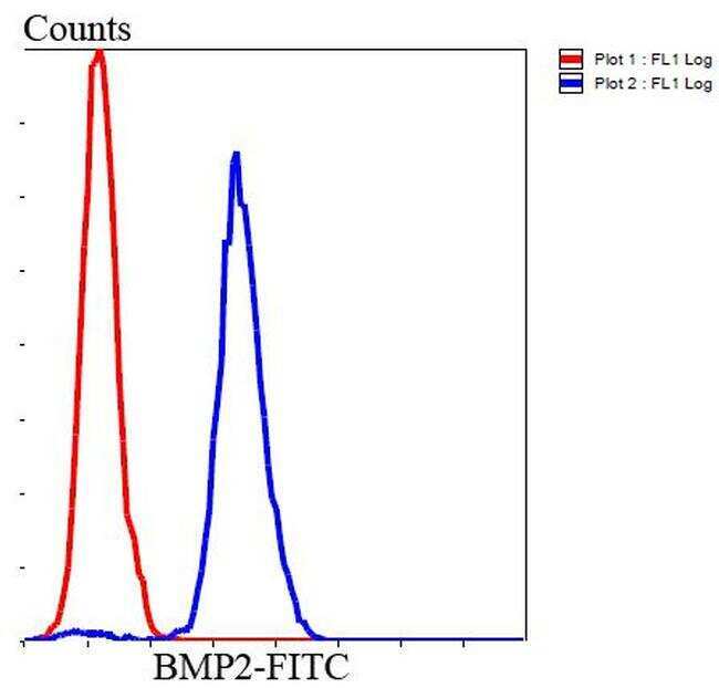
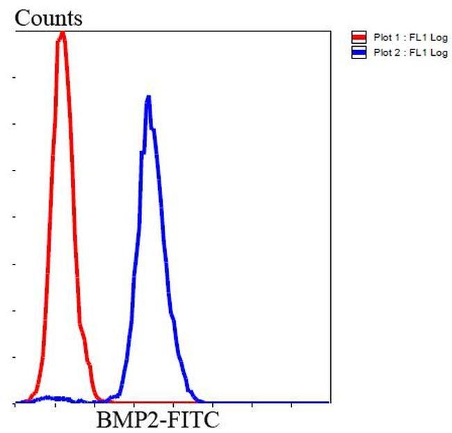
- Flow Cytometric analysis of BMP-2 in Hela cells using a BMP-2 Polyclonal Antibody (Product # PA5-85956) at a dilution of 1:100, as seen in blue compared with an unlabelled control (cells without incubation with primary antibody; red). Goat anti rabbit IgG (FITC) was used as the secondary antibody.
- Submitted by
- Invitrogen Antibodies (provider)
- Main image
- Experimental details
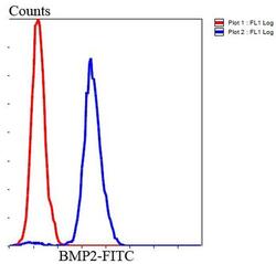
- Flow Cytometric analysis of BMP-2 in Hela cells using a BMP-2 Polyclonal Antibody (Product # PA5-85956) at a dilution of 1:100, as seen in blue compared with an unlabelled control (cells without incubation with primary antibody; red). Goat anti rabbit IgG (FITC) was used as the secondary antibody.
Supportive validation
- Submitted by
- Invitrogen Antibodies (provider)
- Main image
- Experimental details
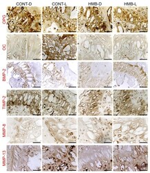
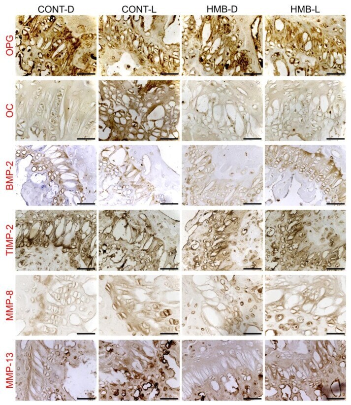
- Figure 5 Representative images of the immunohistochemical reactions for osteoprotegerin (OPG), osteocalcin (OC), bone morphogenetic protein 2 (BMP-2), tissue inhibitor of metalloproteinases 2 (TIMP-2), matrix metalloproteinase 8 (MMP-8) and matrix metalloproteinase 13 (MMP-13) in the growth plate cartilage of femora from pregnant female controls (not receiving HMB) at delivery (CONT-D) or after the lactation period (CONT-L) and from pregnant HMB females (receiving HMB at a dose of 0.02 g/kg b.w. during the middle trimester of pregnancy) at delivery (HMB-D) or after the lactation period (HMB-L). All the scale bars represent 40 um.
- Submitted by
- Invitrogen Antibodies (provider)
- Main image
- Experimental details
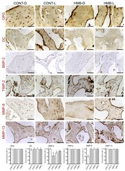
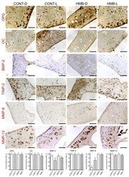
- Figure 6 Representative images of the immunohistochemical reactions and the percent of immunoreactive cells for osteoprotegerin (OPG), osteocalcin (OC), bone morphogenetic protein 2 (BMP-2), tissue inhibitor of metalloproteinases 2 (TIMP-2), matrix metalloproteinase 8 (MMP-8) and matrix metalloproteinase 13 (MMP-13) in the trabecular bone of femora from pregnant female controls (not receiving HMB) at delivery (CONT-D) or after the lactation period (CONT-L) and from pregnant HMB females (receiving HMB at a dose of 0.02 g/kg b.w. during the middle trimester of pregnancy) at delivery (HMB-D) or after the lactation period (HMB-L). All the scale bars represent 40 um. Bar plots show the percentage of immunoreactive osteocytes in the trabecular bone in each group.
- Submitted by
- Invitrogen Antibodies (provider)
- Main image
- Experimental details
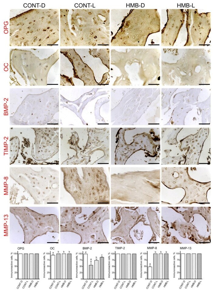
- Figure 7 Representative images of the immunohistochemical reactions and the percent of immunoreactive cells for osteoprotegerin (OPG), osteocalcin (OC), bone morphogenetic protein 2 (BMP-2), tissue inhibitor of metalloproteinases 2 (TIMP-2), matrix metalloproteinase 8 (MMP-8) and matrix metalloproteinase 13 (MMP-13) in the compact bone of femora from pregnant female controls (not receiving HMB) at delivery (CONT-D) or after the lactation period (CONT-L) and from pregnant HMB females (receiving HMB at a dose of 0.02 g/kg b.w. during the middle trimester of pregnancy) at delivery (HMB-D) or after the lactation period (HMB-L). All the scale bars represent 40 um. Bar plots show the percentage of immunoreactive osteocytes in the trabecular bone in each group.
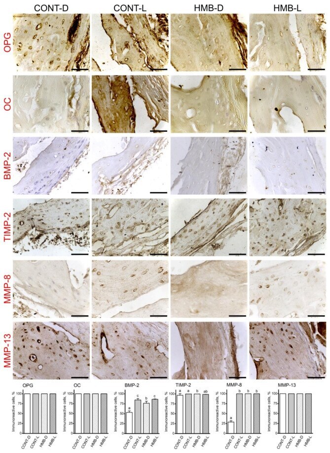
- Submitted by
- Invitrogen Antibodies (provider)
- Main image
- Experimental details
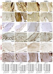
- Figure 8 Representative images of the immunohistochemical reactions and the percent of immunoreactive cells for osteoprotegerin (OPG), osteocalcin (OC), bone morphogenetic protein 2 (BMP-2), tissue inhibitor of metalloproteinases 2 (TIMP-2), matrix metalloproteinase 8 (MMP-8) and matrix metalloproteinase 13 (MMP-13) in the articular cartilage of femora from pregnant female controls (not receiving HMB) at delivery (CONT-D) or after the lactation period (CONT-L) and from pregnant HMB females (receiving HMB at a dose of 0.02 g/kg b.w. during the middle trimester of pregnancy) at delivery (HMB-D) or after the lactation period (HMB-L). All the scale bars represent 40 um. Bar plots show percentage of immunoreactive chondrocytes in the articular cartilage in each group.
