Explore
 Validate
 Learn
Learn Western blot
 Immunocytochemistry
Antibody data
- Antibody Data
- Antigen structure
- References [4]
- Comments [0]
- Validations
- Immunocytochemistry [1]
Submit
Validation data
Reference
Comment
Report error
- Product number
- HPA001901 - Provider product page

- Provider
- Atlas Antibodies
- Proper citation
- Atlas Antibodies Cat#HPA001901, RRID:AB_1078864
- Product name
- Anti-FGB
- Antibody type
- Polyclonal
- Description
- Polyclonal Antibody against Human FGB, Gene description: fibrinogen beta chain, Validated applications: ICC, IHC, WB, Uniprot ID: P02675, Storage: Store at +4°C for short term storage. Long time storage is recommended at -20°C.
- Reactivity
- Human
- Host
- Rabbit
- Conjugate
- Unconjugated
- Isotype
- IgG
- Vial size
- 100 µl
- Concentration
- 0.1 mg/ml
- Storage
- Store at +4°C for short term storage. Long time storage is recommended at -20°C.
- Handling
- The antibody solution should be gently mixed before use.
Submitted references Brain proteome profiling implicates the complement and coagulation cascade in multiple system atrophy brain pathology
A High-throughput Bead-based Affinity Assay Enables Analysis of Genital Protein Signatures in Women At Risk of HIV Infection
Profiling post-centrifugation delay of serum and plasma with antibody bead arrays
Variance decomposition of protein profiles from antibody arrays using a longitudinal twin model
Rydbirk R, Østergaard O, Folke J, Hempel C, DellaValle B, Andresen T, Løkkegaard A, Hejl A, Bode M, Blaabjerg M, Møller M, Danielsen E, Salvesen L, Starhof C, Bech S, Winge K, Rungby J, Pakkenberg B, Brudek T, Olsen J, Aznar S
Cellular and Molecular Life Sciences 2022;79(6)
Cellular and Molecular Life Sciences 2022;79(6)
A High-throughput Bead-based Affinity Assay Enables Analysis of Genital Protein Signatures in Women At Risk of HIV Infection
Månberg A, Bradley F, Qundos U, Guthrie B, Birse K, Noël-Romas L, Lindskog C, Bosire R, Kiarie J, Farquhar C, Burgener A, Nilsson P, Broliden K
Molecular & Cellular Proteomics 2019;18(3):461-476
Molecular & Cellular Proteomics 2019;18(3):461-476
Profiling post-centrifugation delay of serum and plasma with antibody bead arrays
Qundos U, Hong M, Tybring G, Divers M, Odeberg J, Uhlen M, Nilsson P, Schwenk J
Journal of Proteomics 2013;95
Journal of Proteomics 2013;95
Variance decomposition of protein profiles from antibody arrays using a longitudinal twin model
Kato B, Nicholson G, Neiman M, Rantalainen M, Holmes C, Barrett A, Uhlén M, Nilsson P, Spector T, Schwenk J
Proteome Science 2011;9(1):73
Proteome Science 2011;9(1):73
No comments: Submit comment
Supportive validation
- Submitted by
- Atlas Antibodies (provider)
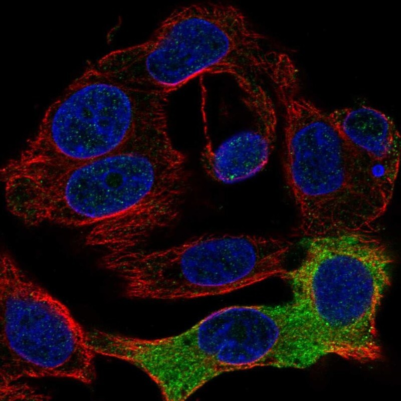
- Main image
- Experimental details
- Immunofluorescent staining of human cell line Hep G2 shows localization to endoplasmic reticulum.
- Sample type
- Human