 Explore
 Validate
 Learn
Learn Western blot
Antibody data
- Antibody Data
- Antigen structure
- References [3]
- Comments [0]
- Validations
- Western blot [2]
Submit
Validation data
Reference
Comment
Report error
- Product number
- AF775 - Provider product page

- Provider
- R&D Systems
- Product name
- Mouse IGFBP-3 Antibody
- Antibody type
- Polyclonal
- Description
- Immunogen affinity purified. Detects mouse IGFBP-3 in direct ELISAs and Western blots. In direct ELISAs, approximately 5% cross-reactivity with recombinant human (rh) IGFBP-3 is observed and less than 1% cross-reactivity with rhIGFBP-1, rhIGFBP-2, rhIGFBP-4, rhIGFBP-5, rhIGFBP-6, and recombinant mouse IGFBP-6 is observed.
- Reactivity
- Mouse
- Host
- Goat
- Conjugate
- Unconjugated
- Antigen sequence
P47878- Isotype
- IgG
- Vial size
- 100 ug
- Concentration
- LYOPH
- Storage
- Use a manual defrost freezer and avoid repeated freeze-thaw cycles. 12 months from date of receipt, -20 to -70 °C as supplied. 1 month, 2 to 8 °C under sterile conditions after reconstitution. 6 months, -20 to -70 °C under sterile conditions after reconstitution.
Submitted references Amyloid beta-mediated epigenetic alteration of insulin-like growth factor binding protein 3 controls cell survival in Alzheimer's disease.
Bi-compartmental communication contributes to the opposite proliferative behavior of Notch1-deficient hair follicle and epidermal keratinocytes.
Loss of SPARC-mediated VEGFR-1 suppression after injury reveals a novel antiangiogenic activity of VEGF-A.
Sung HY, Choi EN, Lyu D, Mook-Jung I, Ahn JH
PloS one 2014;9(6):e99047
PloS one 2014;9(6):e99047
Bi-compartmental communication contributes to the opposite proliferative behavior of Notch1-deficient hair follicle and epidermal keratinocytes.
Lee J, Basak JM, Demehri S, Kopan R
Development (Cambridge, England) 2007 Aug;134(15):2795-806
Development (Cambridge, England) 2007 Aug;134(15):2795-806
Loss of SPARC-mediated VEGFR-1 suppression after injury reveals a novel antiangiogenic activity of VEGF-A.
Nozaki M, Sakurai E, Raisler BJ, Baffi JZ, Witta J, Ogura Y, Brekken RA, Sage EH, Ambati BK, Ambati J
The Journal of clinical investigation 2006 Feb;116(2):422-9
The Journal of clinical investigation 2006 Feb;116(2):422-9
No comments: Submit comment
Supportive validation
- Submitted by
- R&D Systems (provider)
- Main image
- Experimental details
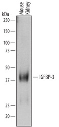
- Detection of Mouse IGFBP-3 by Western Blot. Western blot shows lysates of mouse kidney tissue. PVDF membrane was probed with 0.2 µg/mL of Goat Anti-Mouse IGFBP-3 Antigen Affinity-purified Polyclonal Antibody (Catalog # AF775) followed by HRP-conjugated Anti-Goat IgG Secondary Antibody (Catalog # HAF109). A specific band was detected for IGFBP-3 at approximately 40 kDa (as indicated). This experiment was conducted under reducing conditions and using Immunoblot Buffer Group 1.

- Submitted by
- R&D Systems (provider)
- Main image

- Experimental details
- Detection of Mouse IGFBP-3 by Simple WesternTM. Simple Western lane view shows lysates of mouse kidney tissue, loaded at 0.2 mg/mL. A specific band was detected for IGFBP-3 at approximately 41 kDa (as indicated) using 10 µg/mL of Goat Anti-Mouse IGFBP-3 Antigen Affinity-purified Polyclonal Antibody (Catalog # AF775) followed by 1:50 dilution of HRP-conjugated Anti-Goat IgG Secondary Antibody (Catalog # HAF109). This experiment was conducted under reducing conditions and using the 12-230 kDa separation system.
