Explore
 Validate
 Learn
Learn Western blot
Antibody data
- Antibody Data
- Antigen structure
- References [0]
- Comments [0]
- Validations
- Western blot [3]
- Immunohistochemistry [1]
Submit
Validation data
Reference
Comment
Report error
- Product number
- PA5-27190 - Provider product page

- Provider
- Invitrogen Antibodies
- Product name
- IGFBP3 Polyclonal Antibody
- Antibody type
- Polyclonal
- Antigen
- Recombinant protein fragment
- Description
- Recommended positive controls: 293T, A431, Jurkat, Raji, mouse plasma. Predicted reactivity: Mouse (89%), Rat (88%), Pig (83%), Rhesus Monkey (99%), Bovine (84%). Store product as a concentrated solution. Centrifuge briefly prior to opening the vial.
- Reactivity
- Human, Mouse, Rat
- Host
- Rabbit
- Isotype
- IgG
- Vial size
- 100 µL
- Concentration
- 1 mg/mL
- Storage
- Store at 4°C short term. For long term storage, store at -20°C, avoiding freeze/thaw cycles.
No comments: Submit comment
Supportive validation
- Submitted by
- Invitrogen Antibodies (provider)
- Main image

- Experimental details
- Western blot analysis of IGFBP3 using 30 µg of 293T lysate. Samples were loaded onto a 12% SDS-PAGE gel and probed with an IGFBP3 polyclonal antibody (Product # PA5-27190) at a dilution of 1:1000.

- Submitted by
- Invitrogen Antibodies (provider)
- Main image

- Experimental details
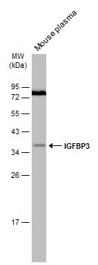
- Western blot analysis of IGFBP3 was performed by separating 30 µg of mouse plasma by 12% SDS-PAGE. Proteins were transferred to a membrane and probed with a IGFBP3 Polyclonal Antibody (Product # PA5-27190) at a dilution of 1:1000. The HRP-conjugated anti-rabbit IgG antibody was used to detect the primary antibody.
- Submitted by
- Invitrogen Antibodies (provider)
- Main image

- Experimental details
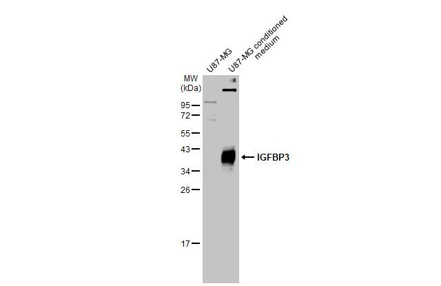
- Western Blot using IGFBP3 Polyclonal Antibody (Product # PA5-27190). U87-MG whole cell extract and conditioned medium (30 µg) were separated by 12% SDS-PAGE, and the membrane was blotted with IGFBP3 Polyclonal Antibody (Product # PA5-27190) diluted at 1:1,000. The HRP-conjugated anti-rabbit IgG antibody was used to detect the primary antibody, and the signal was developed with Trident ECL plus-Enhanced.
Supportive validation
- Submitted by
- Invitrogen Antibodies (provider)
- Main image

- Experimental details
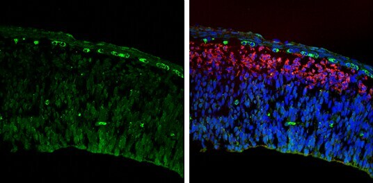
- Immunohistochemistry (Frozen) analysis of IGFBP3 was performed in frozen sectioned E13.5 Rat brain tissue using IGFBP3 Polyclonal Antibody (Product # PA5-27190) at a dilution of 1:250 (Green). Red: beta Tubulin 3/ TUJ1, a mature neuron marker, stained by beta Tubulin 3/ TUJ1 antibody diluted at 1:500. Blue: Fluoroshield with DAPI.