Explore
 Validate
 Learn
Learn Western blot
 Immunocytochemistry
Antibody data
- Antibody Data
- Antigen structure
- References [2]
- Comments [0]
- Validations
- Immunocytochemistry [2]
- Immunohistochemistry [1]
- Other assay [3]
Submit
Validation data
Reference
Comment
Report error
- Product number
- PA5-19963 - Provider product page

- Provider
- Invitrogen Antibodies
- Product name
- Caspase 12 Polyclonal Antibody
- Antibody type
- Polyclonal
- Antigen
- Synthetic peptide
- Description
- A suggested positive control is mouse brain tissue lysate. PA5-19963 can be used with blocking peptide PEP-0088.
- Reactivity
- Human, Mouse, Rat
- Host
- Rabbit
- Isotype
- IgG
- Vial size
- 100 μg
- Concentration
- 1 mg/mL
- Storage
- Maintain refrigerated at 2-8°C for up to 3 months. For long term storage store at -20°C
Submitted references Inhibition of Autophagy Prevents Panax Notoginseng Saponins (PNS) Protection on Cardiac Myocytes Against Endoplasmic Reticulum (ER) Stress-Induced Mitochondrial Injury, Ca(2+) Homeostasis and Associated Apoptosis.
Diazoxide inhibits of ER stress‑mediated apoptosis during oxygen‑glucose deprivation in vitro and cerebral ischemia‑reperfusion in vivo.
Chen J, Li L, Bai X, Xiao L, Shangguan J, Zhang W, Zhang X, Wang S, Liu G
Frontiers in pharmacology 2021;12:620812
Frontiers in pharmacology 2021;12:620812
Diazoxide inhibits of ER stress‑mediated apoptosis during oxygen‑glucose deprivation in vitro and cerebral ischemia‑reperfusion in vivo.
Lei X, Lei L, Zhang Z, Cheng Y
Molecular medicine reports 2018 Jun;17(6):8039-8046
Molecular medicine reports 2018 Jun;17(6):8039-8046
No comments: Submit comment
Supportive validation
- Submitted by
- Invitrogen Antibodies (provider)
- Main image

- Experimental details
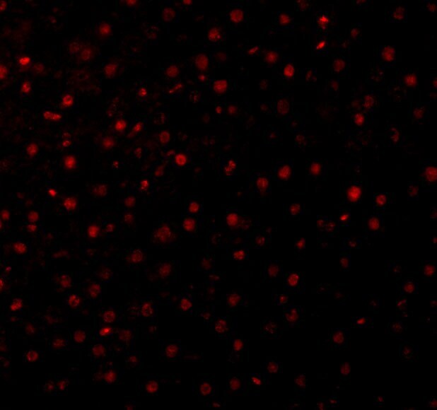
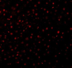
- Immunofluorescence of Caspase-12 in Mouse Liver cells with Caspase 12 Polyclonal Antibody (Product # PA5-19963) at 10 µg/mL.
- Submitted by
- Invitrogen Antibodies (provider)
- Main image
- Experimental details
- Immunofluorescence of Caspase-12 in Mouse Liver cells with Caspase 12 Polyclonal Antibody (Product # PA5-19963) at 10 µg/mL.

Supportive validation
- Submitted by
- Invitrogen Antibodies (provider)
- Main image
- Experimental details
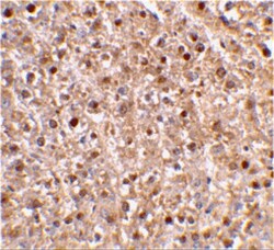
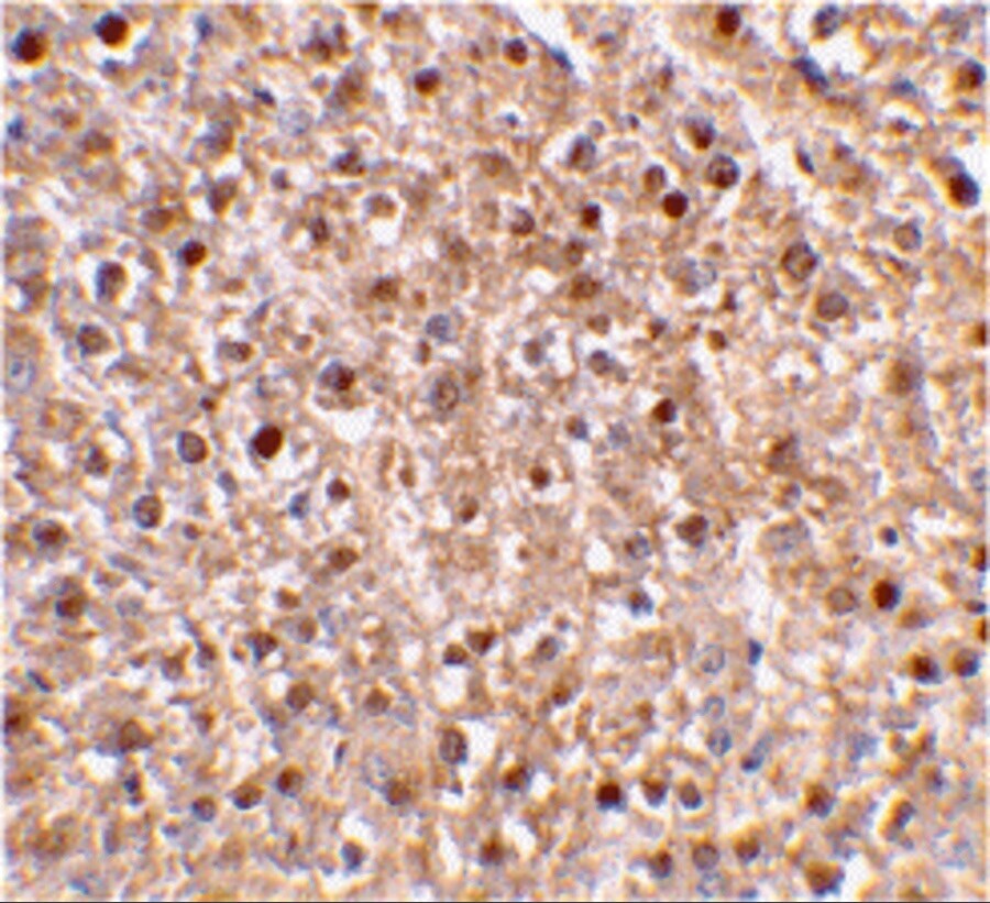
- Immunohistochemical staining of mouse liver tissue using Caspase 12 Polyclonal Antibody (Product # PA5-19963) at 2 µg/mL.
Supportive validation
- Submitted by
- Invitrogen Antibodies (provider)
- Main image
- Experimental details
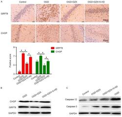
- Figure 2. DZX regulates the expression of CHOP, GRP78, caspase-12 and caspase-3 in OGD-treated hippocampal cells. (A) Immunohistochemical staining demonstrated the distribution of GRP78 and CHOP in hippocampal cells exposed to OGD. (B) Western blot analysis was used to detect the expression of GRP78 and CHOP in hippocampal cells exposed to OGD. (C) Protein expression levels of caspase-12 and caspase-3 in hippocampal cells exposed to OGD were detected using western blotting. Data are expressed as the mean +- standard deviation. *P

- Submitted by
- Invitrogen Antibodies (provider)
- Main image
- Experimental details
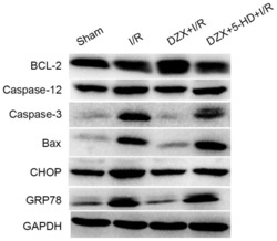
- Figure 5. Pretreatment with DZX modulates the protein expression of Bcl-2, caspase-12, caspase-3, Bax, CHOP and GRP78 in rats subjected to I/R. Western blot analysis was used to detect the protein expression levels of Bcl-2, caspase-12, caspase-3, Bax, CHOP and GRP78 in hippocampal cells isolated from I/R-treated rats. Rats were treated with DZX with or without 5-HD and subjected to cerebral ischemia for 2 h followed by 12 h of reperfusion. Sham rats were not subjected to cerebral ischemia. DZX, diazoxide; Bcl-2, B-cell lymphoma-2; Bax, Bcl-2-associated X protein; CHOP, C/EBP homologous protein; GRP78, 78 kDa glucose-regulated protein; I/R, ischemia/reperfusion; 5-HD, 5-hydroxydecanoate.

- Submitted by
- Invitrogen Antibodies (provider)
- Main image

- Experimental details
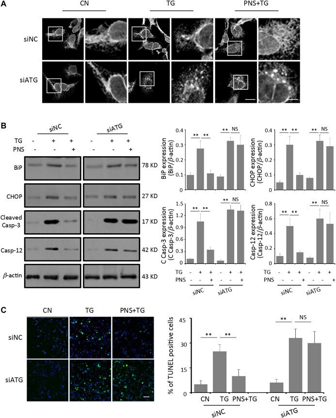
- FIGURE 5 Inhibition of autophagy abolishes PNS protection on TG-induced ER stress response and associated apoptosis. (A) SiNC or siATG7 transfected H9c2 cells, either untreated (CN group) or pretreated with 40 mug/ml PNS for 12 h before addition of 1 muM TG (TG group or PNS plus TG group) for 12 h were immunofluorescenced with the primary anti-calnexin antibody and imaged by a laser scanning confocal microscopy. Scale bar: 30 mum; in box: 10 mum. (B) SiNC or siATG7 transfected H9c2 cells, treated as in A, were immunoblotted with the antibodies to BiP, CHOP, Cleaved Caspase-3 and Caspase-12 as well as beta -actin. Bands were quantified relative to beta -actin by densitometry (Mean +- SEM; ** p < 0.01 relative to CN group, or indicated group). (C) SiNC or siATG7 transfected H9c2 cells, treated as in A, were stained with TUNEL and imaged by a laser scanning confocal microscopy. Scale bar: 200 mum. Bar graph shows the percentage of TUNEL positive cells (Mean +- SEM; 200-250 cells; ** p < 0.01 relative to CN group, or indicated group).