 Explore
 Validate
 Learn
Learn Western blot
 Immunoprecipitation
Antibody data
- Antibody Data
- Antigen structure
- References [7]
- Comments [0]
- Validations
- Western blot [3]
- Immunohistochemistry [1]
Submit
Validation data
Reference
Comment
Report error
- Product number
- AF1014 - Provider product page

- Provider
- Novus Biologicals
- Product name
- Goat Polyclonal Cathepsin D Antibody
- Antibody type
- Polyclonal
- Description
- Antigen Affinity-purified. Detects human Cathepsin D in direct ELISAs and Western blots. In direct ELISAs, approximately 20% cross-reactivity with recombinant mouse (rm) Cathepsin D is observed.
- Reactivity
- Human
- Host
- Goat
- Conjugate
- Unconjugated
- Isotype
- IgG
- Vial size
- 100 ug
- Concentration
- LYOPH
- Storage
- Use a manual defrost freezer and avoid repeated freeze-thaw cycles. 12 months from date of receipt, -20 to -70 degreesC as supplied. 1 month, 2 to 8 degreesC under sterile conditions after reconstitution. 6 months, -20 to -70 degreesC under sterile conditions after reconstitution.
Submitted references Identification of PIKfyve kinase as a target in multiple myeloma.
Visualizing the cellular route of entry of a cystine-knot peptide with Xfect transfection reagent by electron microscopy.
HSP90 inhibition targets autophagy and induces a CASP9-dependent resistance mechanism in NSCLC.
Amphiphilic star PEG-Camptothecin conjugates for intracellular targeting.
The Apaf-1-binding protein Aven is cleaved by Cathepsin D to unleash its anti-apoptotic potential.
IFN-gamma regulation of vacuolar pH, cathepsin D processing and autophagy in mammary epithelial cells.
Clathrin is a key regulator of basolateral polarity.
de Campos CB, Zhu YX, Sepetov N, Romanov S, Bruins LA, Shi CX, Stein CK, Petit JL, Polito AN, Sharik ME, Meermeier EW, Ahmann GJ, Armenta IDL, Kruse J, Bergsagel PL, Chesi M, Meurice N, Braggio E, Stewart AK
Haematologica 2020 Jun;105(6):1641-1649
Haematologica 2020 Jun;105(6):1641-1649
Visualizing the cellular route of entry of a cystine-knot peptide with Xfect transfection reagent by electron microscopy.
Gao X, De Mazière A, Iaea DB, Arthur CP, Klumperman J, Ciferri C, Hannoush RN
Scientific reports 2019 May 6;9(1):6907
Scientific reports 2019 May 6;9(1):6907
HSP90 inhibition targets autophagy and induces a CASP9-dependent resistance mechanism in NSCLC.
Han J, Goldstein LA, Hou W, Chatterjee S, Burns TF, Rabinowich H
Autophagy 2018;14(6):958-971
Autophagy 2018;14(6):958-971
Amphiphilic star PEG-Camptothecin conjugates for intracellular targeting.
Omar R, Bardoogo YL, Corem-Salkmon E, Mizrahi B
Journal of controlled release : official journal of the Controlled Release Society 2017 Jul 10;257:76-83
Journal of controlled release : official journal of the Controlled Release Society 2017 Jul 10;257:76-83
The Apaf-1-binding protein Aven is cleaved by Cathepsin D to unleash its anti-apoptotic potential.
Melzer IM, Fernández SB, Bösser S, Lohrig K, Lewandrowski U, Wolters D, Kehrloesser S, Brezniceanu ML, Theos AC, Irusta PM, Impens F, Gevaert K, Zörnig M
Cell death and differentiation 2012 Sep;19(9):1435-45
Cell death and differentiation 2012 Sep;19(9):1435-45
IFN-gamma regulation of vacuolar pH, cathepsin D processing and autophagy in mammary epithelial cells.
Khalkhali-Ellis Z, Abbott DE, Bailey CM, Goossens W, Margaryan NV, Gluck SL, Reuveni M, Hendrix MJ
Journal of cellular biochemistry 2008 Sep 1;105(1):208-18
Journal of cellular biochemistry 2008 Sep 1;105(1):208-18
Clathrin is a key regulator of basolateral polarity.
Deborde S, Perret E, Gravotta D, Deora A, Salvarezza S, Schreiner R, Rodriguez-Boulan E
Nature 2008 Apr 10;452(7188):719-23
Nature 2008 Apr 10;452(7188):719-23
No comments: Submit comment
Supportive validation
- Submitted by
- Novus Biologicals (provider)
- Main image

- Experimental details
- Detection of Human Cathepsin D by Simple Western<sup abp="263">TM. Simple Western lane view shows lysates of K562 human chronic myelogenous leukemia cell line and MCF-7 human breast cancer cell line, loaded at 0.2 mg/mL. Specific bands were detected for Procathepsin D at approximately 55 kDa and Cathepsin D heavy chain at approximately 37 kDa (as indicated) using 50 µg/mL of Goat Anti-Human Cathepsin D Antigen Affinity-purified Polyclonal Antibody (Catalog # AF1014) followed by 1:50 dilution of HRP-conjugated Anti-Goat IgG Secondary Antibody (Catalog # HAF109). This experiment was conducted under reducing conditions and using the 12-230 kDa separation system. Non-specific interaction with the 230 kDa Simple Western standard may be seen with this antibody.

- Submitted by
- Novus Biologicals (provider)
- Main image

- Experimental details
- Detection of Human Cathepsin D by Western Blot. Western blot shows lysates of K562 human chronic myelogenous leukemia cell line. PVDF membrane was probed with 1 µg/mL of Goat Anti-Human Cathepsin D Antigen Affinity-purified Polyclonal Antibody (Catalog # AF1014) followed by HRP-conjugated Anti-Goat IgG Secondary Antibody (Catalog # HAF019). Specific bands were detected for Procathepsin D at approximately 45 kDa and Cathepsin D heavy chain 28 kDa (as indicated). This experiment was conducted under reducing conditions and using Immunoblot Buffer Group 1.

- Submitted by
- Novus Biologicals (provider)
- Main image

- Experimental details
- Detection of Human Cathepsin D by Western Blot. Western blot shows lysates of PANC-1 human pancreatic carcinoma cell line and MCF-7 human breast cancer cell line. PVDF membrane was probed with 1 µg/mL of Goat Anti-Human Cathepsin D Antigen Affinity-purified Polyclonal Antibody (Catalog # AF1014) followed by HRP-conjugated Anti-Goat IgG Secondary Antibody (Catalog # HAF017). Specific bands were detected for Procathepsin D at approximately 45 kDa and Cathepsin D heavy chain 28 kDa (as indicated). This experiment was conducted under reducing conditions and using Immunoblot Buffer Group 1.

Supportive validation
- Submitted by
- Novus Biologicals (provider)
- Main image
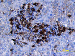
- Experimental details
- Cathepsin D in Human Lung Cancer Tissue. Cathepsin D was detected in immersion fixed paraffin-embedded sections of human lung cancer tissue using Goat Anti-Human Cathepsin D Antigen Affinity-purified Polyclonal Antibody (Catalog # AF1014) at 15 µg/mL overnight at 4 °C. Tissue was stained using the Anti-Goat HRP-DAB Cell & Tissue Staining Kit (brown; Catalog # CTS008) and counterstained with hematoxylin (blue). View our protocol for Chromogenic IHC Staining of Paraffin-embedded Tissue Sections.
