Explore
 Validate
 Learn
Learn Western blot
 Immunocytochemistry
Antibody data
- Antibody Data
- Antigen structure
- References [5]
- Comments [0]
- Validations
- Western blot [2]
- Immunohistochemistry [1]
Submit
Validation data
Reference
Comment
Report error
- Product number
- GTX62063 - Provider product page

- Provider
- GeneTex
- Proper citation
- GeneTex Cat#GTX62063, RRID:AB_10622576
- Product name
- Cathepsin D antibody [EPR3057Y]
- Antibody type
- Monoclonal
- Reactivity
- Human, Mouse
- Host
- Rabbit
- Storage
- Store at -20¢XC. Stable for 12 months at -20¢XC
Submitted references K63 linked ubiquitin chain formation is a signal for HIF1A degradation by Chaperone-Mediated Autophagy.
PIKfyve inhibition interferes with phagosome and endosome maturation in macrophages.
Lysosomal membrane permeabilization: carbon nanohorn-induced reactive oxygen species generation and toxicity by this neglected mechanism.
Proteomic analysis of rhein-induced cyt: ER stress mediates cell death in breast cancer cells.
Lysosomal Cathepsin D contributes to cell death during adipocyte hypertrophy.
Ferreira JV, Soares AR, Ramalho JS, Pereira P, Girao H
Scientific reports 2015 May 11;5:10210
Scientific reports 2015 May 11;5:10210
PIKfyve inhibition interferes with phagosome and endosome maturation in macrophages.
Kim GH, Dayam RM, Prashar A, Terebiznik M, Botelho RJ
Traffic (Copenhagen, Denmark) 2014 Oct;15(10):1143-63
Traffic (Copenhagen, Denmark) 2014 Oct;15(10):1143-63
Lysosomal membrane permeabilization: carbon nanohorn-induced reactive oxygen species generation and toxicity by this neglected mechanism.
Yang M, Zhang M, Tahara Y, Chechetka S, Miyako E, Iijima S, Yudasaka M
Toxicology and applied pharmacology 2014 Oct 1;280(1):117-26
Toxicology and applied pharmacology 2014 Oct 1;280(1):117-26
Proteomic analysis of rhein-induced cyt: ER stress mediates cell death in breast cancer cells.
Huang HJ, Lin CC, Chou HC, Chen YW, Lin ST, Lin YC, Lin DY, Lyu KW, Chan HL
Molecular bioSystems 2014 Dec;10(12):3086-100
Molecular bioSystems 2014 Dec;10(12):3086-100
Lysosomal Cathepsin D contributes to cell death during adipocyte hypertrophy.
Eguchi A, Feldstein AE
Adipocyte 2013 Jul 1;2(3):170-5
Adipocyte 2013 Jul 1;2(3):170-5
No comments: Submit comment
Supportive validation
- Submitted by
- GeneTex (provider)
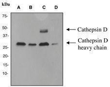
- Main image
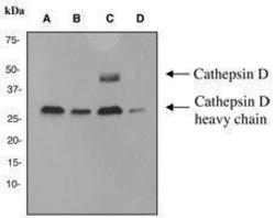
- Experimental details
- A.Western blot analysis on (A) MCF-7 (B) A431 (C) SKBR3 and (D) HepG2 cell lysates using anti-Cathepsin D RabMAb (cat. # GTX62063) dilution 1:10000.
- Validation comment
- WB
- Submitted by
- GeneTex (provider)
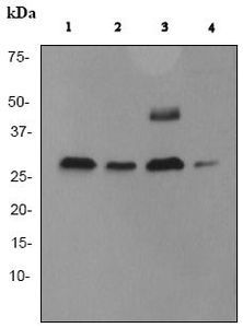
- Main image

- Experimental details
- WB analysis of (1) MCF-7, (2) A431, (3) SKBR3 and (4) HepG2 lysates (10 £gg per lane) using Cathepsin D antibody [EPR3057Y] at a dilution of 1:10,000.
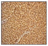
Supportive validation
- Submitted by
- GeneTex (provider)
- Main image
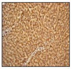
- Experimental details
- IHC-P analysis of human liver tissue using Cathepsin D antibody [EPR3057Y] at a dilution of 1:500.