Explore
 Validate
 Learn
Learn Western blot
 Immunocytochemistry
Antibody data
- Antibody Data
- Antigen structure
- References [1]
- Comments [0]
- Validations
- Western blot [1]
Submit
Validation data
Reference
Comment
Report error
- Product number
- PB9854 - Provider product page

- Provider
- Boster Biological Technology
- Product name
- Anti-Cathepsin D/CTSD Antibody Picoband™
- Antibody type
- Polyclonal
- Description
- Polyclonal antibody for Cathepsin D/CTSD detection. Host: Rabbit.Size: 100μg/vial. Tested applications: WB, IHC-P, ICC/IF, FCM. Reactive species: Human. Cathepsin D/CTSD information: Molecular Weight: 44552 MW; Subcellular Localization: Lysosome. Melanosome. Secreted, extracellular space. Identified by mass spectrometry in melanosome fractions from stage I to stage IV. In aortic samples, detected as an extracellular protein loosely bound to the matrix (PubMed:20551380); Tissue Specificity: Expressed in the aorta extrcellular space (at protein level).
- Reactivity
- Human
- Host
- Rabbit
- Vial size
- 100μg/vial
- Concentration
- Add 0.2ml of distilled water will yield a concentration of 500ug/ml.
- Storage
- At -20°C for one year. After reconstitution, at 4°C for one month. It can also be aliquoted and stored frozen at -20°C for a longer time. Avoid repeated freezing and thawing.
- Handling
- Add 0.2ml of distilled water will yield a concentration of 500ug/ml.
Submitted references Identification of chemoresistance-related cell-surface glycoproteins in leukemia cells and functional validation of candidate glycoproteins.
Sun Z, Dong J, Zhang S, Hu Z, Cheng K, Li K, Xu B, Ye M, Nie Y, Fan D, Zou H
Journal of proteome research 2014 Mar 7;13(3):1593-601
Journal of proteome research 2014 Mar 7;13(3):1593-601
No comments: Submit comment
Supportive validation
- Submitted by
- Boster Biological Technology (provider)
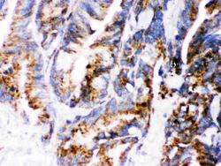
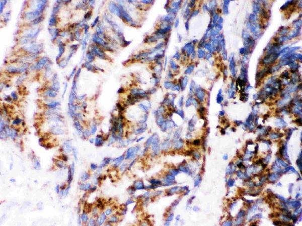
- Main image
- Experimental details
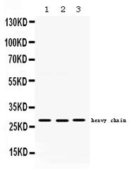
- Western blot analysis of Cathepsin D using anti-Cathepsin D antibody (PB9854). Electrophoresis was performed on a 5-20% SDS-PAGE gel at 70V (Stacking gel) / 90V (Resolving gel) for 2-3 hours. The sample well of each lane was loaded with 50ug of sample under reducing conditions. Lane 1: HEPG2 whole cell lysates, Lane 2: A549 whole cell lysates, Lane 3: PANC1 whole cell lysates. After Electrophoresis, proteins were transferred to a Nitrocellulose membrane at 150mA for 50-90 minutes. Blocked the membrane with 5% Non-fat Milk/ TBS for 1.5 hour at RT. The membrane was incubated with rabbit anti-Cathepsin D antigen affinity purified polyclonal antibody (Catalog # PB9854) at 0.5 μg/mL overnight at 4°C, then washed with TBS-0.1%Tween 3 times with 5 minutes each and probed with a goat anti-rabbit IgG-HRP secondary antibody at a dilution of 1:10000 for 1.5 hour at RT. The signal is developed using an Enhanced Chemiluminescent detection (ECL) kit (Catalog # EK1002) with Tanon 5200 system. A specific band was detected for Cathepsin D at approximately 28KD. The expected band size for Cathepsin D is at 28KD.
- Additional image