 Explore
 Validate
 Learn
LearnNBP1-04278
antibody from Novus Biologicals
Targeting: CTSD
CLN10, CPSD
 Western blot
 ELISA Immunocytochemistry Immunohistochemistry Flow cytometry Immunoelectron microscopy
Antibody data
- Antibody Data
- Antigen structure
- References [4]
- Comments [0]
- Validations
- Western blot [2]
- Immunohistochemistry [2]
- Flow cytometry [1]
Submit
Validation data
Reference
Comment
Report error
- Product number
- NBP1-04278 - Provider product page

- Provider
- Novus Biologicals
- Proper citation
- Novus Cat#NBP1-04278, RRID:AB_1520484
- Product name
- Mouse Monoclonal Cathepsin D Antibody
- Antibody type
- Monoclonal
- Description
- Protein G purified.
- Reactivity
- Human
- Host
- Mouse
- Isotype
- IgG
- Vial size
- 0.1 ml
- Concentration
- 1.0 mg/ml
- Storage
- Store at 4C short term. Aliquot and store at -20C long term. Avoid freeze-thaw cycles.
Submitted references Naturally Occurring Variants in LRP1 (Low-Density Lipoprotein Receptor-Related Protein 1) Affect HDL (High-Density Lipoprotein) Metabolism Through ABCA1 (ATP-Binding Cassette A1) and SR-B1 (Scavenger Receptor Class B Type 1) in Humans.
Natural history of Helicobacter pylori VacA toxin in human gastric epithelium in vivo: vacuoles and beyond.
Mieap, a p53-inducible protein, controls mitochondrial quality by repairing or eliminating unhealthy mitochondria.
Possible existence of lysosome-like organella within mitochondria and its role in mitochondrial quality control.
Oldoni F, van Capelleveen JC, Dalila N, Wolters JC, Heeren J, Sinke RJ, Hui DY, Dallinga-Thie GM, Frikke-Schmidt R, Hovingh KG, van de Sluis B, Tybjærg-Hansen A, Kuivenhoven JA
Arteriosclerosis, thrombosis, and vascular biology 2018 Jul;38(7):1440-1453
Arteriosclerosis, thrombosis, and vascular biology 2018 Jul;38(7):1440-1453
Natural history of Helicobacter pylori VacA toxin in human gastric epithelium in vivo: vacuoles and beyond.
Necchi V, Sommi P, Vanoli A, Fiocca R, Ricci V, Solcia E
Scientific reports 2017 Nov 6;7(1):14526
Scientific reports 2017 Nov 6;7(1):14526
Mieap, a p53-inducible protein, controls mitochondrial quality by repairing or eliminating unhealthy mitochondria.
Kitamura N, Nakamura Y, Miyamoto Y, Miyamoto T, Kabu K, Yoshida M, Futamura M, Ichinose S, Arakawa H
PloS one 2011 Jan 17;6(1):e16060
PloS one 2011 Jan 17;6(1):e16060
Possible existence of lysosome-like organella within mitochondria and its role in mitochondrial quality control.
Miyamoto Y, Kitamura N, Nakamura Y, Futamura M, Miyamoto T, Yoshida M, Ono M, Ichinose S, Arakawa H
PloS one 2011 Jan 17;6(1):e16054
PloS one 2011 Jan 17;6(1):e16054
No comments: Submit comment
Supportive validation
- Submitted by
- Novus Biologicals (provider)
- Main image
- Experimental details
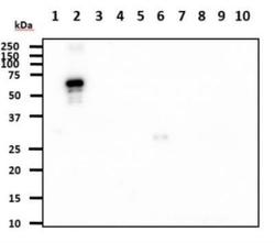
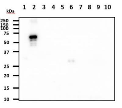
- Western Blot: Cathepsin D Antibody (4G2) [NBP1-04278] - The recombinant proteins (100ng) were resolved by SDSPAGE, transferred to PVDF membrane and probed with anti human Cathepsin D antibody (1:1000). Proteins were visualized using a goat anti-mouse secondary antibody conjugated to HRP and an ECL detection system. Lane 1. : Recombinant Human CTSB Lane 2. : Recombinant Human CTSD Lane 3. : Recombinant Human CTSE Lane 4. : Recombinant Human CTSF Lane 5. : Recombinant Human CTSH Lane 6. : Recombinant Human CTSK Lane 7. : Recombinant Human CTSL Lane 8. : Recombinant Human CTSS Lane 9. : Recombinant Human CTSW Lane 10. : Recombinant Human CTSZ
- Submitted by
- Novus Biologicals (provider)
- Main image
- Experimental details
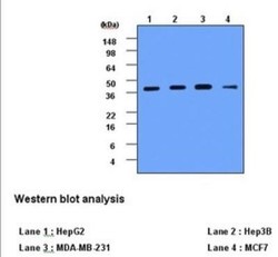
- Western Blot: Cathepsin D Antibody (4G2) [NBP1-04278] - Western Blot: Cathepsin D antibody (clone 4G2)[NBP04278]-Cell lysates of HepG2, Hep3B, MDA-MB-231 and MCF7 (each 20ug) were resolved by SDS-PAGE, transferred to PVDF membrane and probed with anti-human Cathepsin D (1:1000). Proteins were visualized using a goat anti-mouse secondary antibody conjugated to HRP and ECL detection system.

Supportive validation
- Submitted by
- Novus Biologicals (provider)
- Main image
- Experimental details
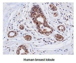
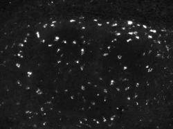
- Immunohistochemistry-Paraffin: Cathepsin D Antibody (4G2) [NBP1-04278] - human Cathepsin D (1:50) for 2 hours at room temperature. Antigen retrieval was performed in 0.1M sodium citrate buffer and detected using Diaminobenzidine (DAB)

- Submitted by
- Novus Biologicals (provider)
- Main image
- Experimental details
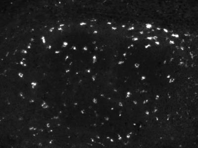
- Immunohistochemistry-Paraffin: Cathepsin D Antibody (4G2) [NBP1-04278] - Cathepsin D 1:50 on tonsil, pH 9 antigen retrieval. IHC-P image submitted by a verified customer review.
Supportive validation
- Submitted by
- Novus Biologicals (provider)
- Main image
- Experimental details
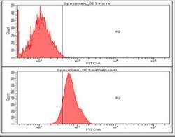
- Flow Cytometry: Cathepsin D Antibody (4G2) [NBP1-04278] - Analysis of Cathepsin D in Hep3B cell line, staining at 2-5ug for 1x10⁶cells. The secondary antibody used goat anti-mouse IgG Alexa fluor 488 conjugate.
