Explore
 Validate
 Learn
Learn Western blot
 Immunohistochemistry
Antibody data
- Antibody Data
- Antigen structure
- References [1]
- Comments [0]
- Validations
- Immunohistochemistry [1]
Submit
Validation data
Reference
Comment
Report error
- Product number
- M01361 - Provider product page

- Provider
- Boster Biological Technology
- Product name
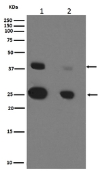
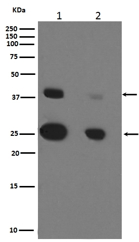
- Anti-Cathepsin D CTSD Rabbit Monoclonal Antibody
- Antibody type
- Monoclonal
- Description
- Monoclonal antibody for Cathepsin D/CTSD detection. Host: Rabbit.Size: 100ug/vial. Tested applications: Flow Cytometry, IP, IF, IHC, ICC, WB. Reactive species: Human, Mouse, Rat Cathepsin D/CTSD information: Molecular Weight: 44552 MW; Subcellular Localization: Lysosome. Melanosome. Secreted, extracellular space. Identified by mass spectrometry in melanosome fractions from stage I to stage IV. In aortic samples, detected as an extracellular protein loosely bound to the matrix (PubMed:20551380); Tissue Specificity: Expressed in the aorta extrcellular space (at protein level).
- Reactivity
- Human, Mouse, Rat
- Host
- Rabbit
- Antibody clone number
- CEF-3
- Vial size
- 100ug/vial
- Concentration
- 0.5-1mg/ml, actual concentration vary by lot. Use suggested dilution ratio to decide dilution procedure.
- Storage
- At -20°C for one year. Avoid repeated freezing and thawing.
Submitted references Identification of chemoresistance-related cell-surface glycoproteins in leukemia cells and functional validation of candidate glycoproteins.
Sun Z, Dong J, Zhang S, Hu Z, Cheng K, Li K, Xu B, Ye M, Nie Y, Fan D, Zou H
Journal of proteome research 2014 Mar 7;13(3):1593-601
Journal of proteome research 2014 Mar 7;13(3):1593-601
No comments: Submit comment
Supportive validation
- Submitted by
- Boster Biological Technology (provider)
- Main image
- Experimental details
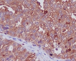
- Immunohistochemical analysis on paraffin-embedded tisf paraffin-embedded human ovarian cancer tissue, using Cathepsin D Antibody(M01361)CTSD was detected isue section. Heat mediated antigen retrieval was performed in citrate buffer (pH6, epitope retrieval solution) for 20 mins. The tissue section was blocked with 10% goat serum. The tissue section was then incubated with 1ug/ml rabbit anti-CTSD Antibody (M01361)overnight at 4
- Additional image