 Explore
 Validate
 Learn
Learn Western blot
 ELISA
ELISAAntibody data
- Antibody Data
- Antigen structure
- References [0]
- Comments [0]
- Validations
- Western blot [2]
- Immunohistochemistry [5]
Submit
Validation data
Reference
Comment
Report error
- Product number
- AM09003PU-S - Provider product page

- Provider
- Acris Antibodies GmbH
- Proper citation
- Acris Antibodies GmbH Cat#AM09003PU-S, RRID:AB_1609688
- Product name
- anti Cathepsin D
- Antibody type
- Monoclonal
- Antigen
- Recombinant human Cathepsin D (21-412 aa) purified from E. coli
- Reactivity
- Human
- Host
- Mouse
- Isotype
- IgG
- Antibody clone number
- 4G2
- Vial size
- 50 µl
- Concentration
- 1.0 mg/ml
No comments: Submit comment
Supportive validation
- Submitted by
- Acris Antibodies GmbH (provider)
- Main image
- Experimental details
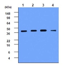
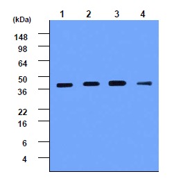
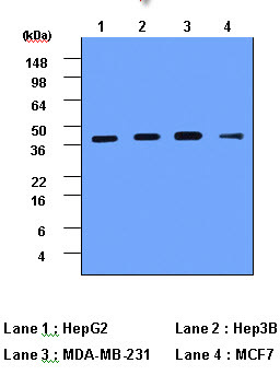
- Western blot analysis: Cell lysates of HepG2 (lane 1), Hep3B (lane 2), MDA-MB-231 (lane 3) and MCF7 (lane 4) -each 20 ug- were resolved by SDS-PAGE, transferred to PVDF membrane and probed with anti-human Cathepsin D (1:1000). Proteins were visualized using a goat anti-mouse secondary antibody conjugated to HRP and ECL detection system.
- Submitted by
- Acris Antibodies GmbH (provider)
- Main image

- Experimental details
- Cell lysates of HepG2, Hep3B, MDA-MB-231 and MCF7 (each 20ug) were resolved by SDS-PAGE, transferred to PVDF membrane and probed with anti-human Cathepsin D (1:1000). Proteins were visualized using a goat anti-mouse secondary antibody conjugated to HRP and ECL detection system.
Supportive validation
- Submitted by
- Acris Antibodies GmbH (provider)
- Main image
- Experimental details
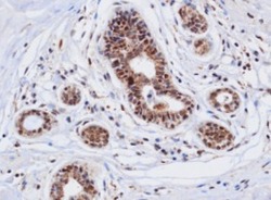
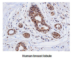
- Immunohistochemistry: Paraffin embedded sections of human breast lobule tissue were incubated with anti-human Cathepsin D (1:50) for 2 hours at room temperature. Antigen retrieval was performed in 0.1 M sodium citrate buffer. Diaminobenzidine (DAB) was used for detection.

- Submitted by
- Acris Antibodies GmbH (provider)
- Main image
- Experimental details
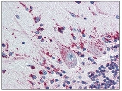
- Immunohistochemistry: AM09003PU-N Cathepsin D antibody staining of Formalin-Fixed, Paraffin-Embedded Human Brain , Cerebellum at 5 µg/ml followed by biotinylated anti-Mouse IgG secondary antibody, Alkaline Phosphatase-Streptavidin and chromogen.

- Submitted by
- Acris Antibodies GmbH (provider)
- Main image
- Experimental details
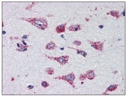
- Immunohistochemistry: AM09003PU-N Cathepsin D antibody staining of Formalin-Fixed, Paraffin-Embedded Human Brain , Cortex at 5 µg/ml followed by biotinylated anti-Mouse IgG secondary antibody, Alkaline Phosphatase-Streptavidin and chromogen.

- Submitted by
- Acris Antibodies GmbH (provider)
- Main image
- Experimental details
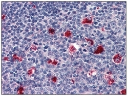
- Immunohistochemistry: AM09003PU-N Cathepsin D antibody staining of Formalin-Fixed, Paraffin-Embedded Human Tonsil at 5 µg/ml followed by biotinylated anti-Mouse IgG secondary antibody, Alkaline Phosphatase-Streptavidin and chromogen.

- Submitted by
- Acris Antibodies GmbH (provider)
- Main image
- Experimental details
- Paraffin embedded sections of human breast lobule tissue were incubated with anti-human Cathepsin D (1:50) for 2 hours at room temperature. Antigen retrieval was performed in 0.1M sodium citrate buffer and detected using Diaminobenzidine (DAB)
