 Explore
 Validate
 Learn
Learn Western blot
Antibody data
- Antibody Data
- Antigen structure
- References [1]
- Comments [0]
- Validations
- Western blot [2]
- Immunohistochemistry [1]
Submit
Validation data
Reference
Comment
Report error
- Product number
- MAB1014 - Provider product page

- Provider
- R&D Systems
- Product name
- Human Cathepsin D Antibody
- Antibody type
- Monoclonal
- Description
- Protein A or G purified from hybridoma culture supernatant. Detects human Cathepsin D in direct ELISAs and Western blots. It recognizes both the pro and mature forms of recombinant human (rh) Cathepsin D. In direct ELISAs, no cross-reactivity with rhCathepsin A, rhCathepsin B, rhCathepsin C, rhCathepsin L, rhCathepsin O, rhCathepsin S, rhCathepsin Z, or recombinant mouse Cathepsin D is observed. In Western blots, 100% cross-reactivity with rhCathepsin E and rmCathepsin D is observed and no cross-reactivity with rhBACE-1 is observed.
- Reactivity
- Human
- Host
- Mouse
- Conjugate
- Unconjugated
- Antigen sequence
P07339- Isotype
- IgG
- Antibody clone number
- 185111
- Vial size
- 500 ug
- Concentration
- LYOPH
- Storage
- Use a manual defrost freezer and avoid repeated freeze-thaw cycles. 12 months from date of receipt, -20 to -70 °C as supplied. 1 month, 2 to 8 °C under sterile conditions after reconstitution. 6 months, -20 to -70 °C under sterile conditions after reconstitution.
Submitted references Biomarker discovery from pancreatic cancer secretome using a differential proteomic approach.
Grønborg M, Kristiansen TZ, Iwahori A, Chang R, Reddy R, Sato N, Molina H, Jensen ON, Hruban RH, Goggins MG, Maitra A, Pandey A
Molecular & cellular proteomics : MCP 2006 Jan;5(1):157-71
Molecular & cellular proteomics : MCP 2006 Jan;5(1):157-71
No comments: Submit comment
Supportive validation
- Submitted by
- R&D Systems (provider)
- Main image

- Experimental details
- Detection of Human Cathepsin D by Western Blot. Western blot shows lysates of PANC-1 human pancreatic carcinoma cell line and MCF-7 human breast cancer cell line. PVDF membrane was probed with 0.2 µg/mL of Mouse Anti-Human Cathepsin D Monoclonal Antibody (Catalog # MAB1014) followed by HRP-conjugated Anti-Mouse IgG Secondary Antibody (Catalog # HAF018). Specific bands were detected for Cathepsin D at approximately 28 and 46 kDa (as indicated). This experiment was conducted under reducing conditions and using Immunoblot Buffer Group 1.

- Submitted by
- R&D Systems (provider)
- Main image

- Experimental details
- Detection of Human Cathepsin D by Simple WesternTM. Simple Western lane view shows lysates of PANC-1 human pancreatic carcinoma cell line and MCF-7 human breast cancer cell line, loaded at 0.2 mg/mL. Specific bands were detected for Cathepsin D at approximately 36 and 52-57 kDa (as indicated) using 2 µg/mL of Mouse Anti-Human Cathepsin D Monoclonal Antibody (Catalog # MAB1014) . This experiment was conducted under reducing conditions and using the 12-230 kDa separation system.

Supportive validation
- Submitted by
- R&D Systems (provider)
- Main image
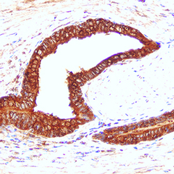
- Experimental details
- Cathepsin D in Human Prostate Cancer Tissue. Cathepsin D was detected in immersion fixed paraffin-embedded sections of human prostate cancer tissue using Mouse Anti-Human Cathepsin D Monoclonal Antibody (Catalog # MAB1014) at 5 µg/mL for 1 hour at room temperature followed by incubation with the Anti-Mouse IgG VisUCyte™ HRP Polymer Antibody (Catalog # VC001). Tissue was stained using DAB (brown) and counterstained with hematoxylin (blue). Specific staining was localized to epithelial cell cytoplasm. View our protocol for IHC Staining with VisUCyte HRP Polymer Detection Reagents.
