 Explore
 Validate
 Learn
Learn Western blot
 Immunoprecipitation
Antibody data
- Antibody Data
- Antigen structure
- References [7]
- Comments [0]
- Validations
- Western blot [1]
- Immunocytochemistry [4]
Submit
Validation data
Reference
Comment
Report error
- Product number
- NB300-525 - Provider product page

- Provider
- Novus Biologicals
- Proper citation
- Novus Cat#NB300-525, RRID:AB_10001514
- Product name
- Mouse Monoclonal ARNT/HIF-1 beta Antibody
- Antibody type
- Monoclonal
- Description
- Immunogen affinity purified. Detects aryl hydrocarbon (Ah) receptor nuclear translocator (ARNT)
- Reactivity
- Human, Mouse, Rat, Simian, Zebrafish
- Host
- Mouse
- Isotype
- IgG
- Vial size
- 100 uL
- Concentration
- 1 mg/ml
- Storage
- Store at -20C. Avoid freeze-thaw cycles.
Submitted references ARNT is a potential direct HIF-1 target gene in human Hep3B hepatocellular carcinoma cells.
A HIF-1α-driven feed-forward loop augments HIF signalling in Hep3B cells by upregulation of ARNT.
The expression level of the transcription factor Aryl hydrocarbon receptor nuclear translocator (ARNT) determines cellular survival after radiation treatment.
CD24 is an effector of HIF-1-driven primary tumor growth and metastasis.
EGFR variant-mediated invasion by enhanced CXCR4 expression through transcriptional and post-translational mechanisms.
Treatment of mice with the Ah receptor agonist and human carcinogen dioxin results in altered numbers and function of hematopoietic stem cells.
Physicochemical and immunocytochemical analysis of the aryl hydrocarbon receptor nuclear translocator: characterization of two monoclonal antibodies to the aryl hydrocarbon receptor nuclear translocator.
Mandl M, Depping R
Cancer cell international 2017;17:77
Cancer cell international 2017;17:77
A HIF-1α-driven feed-forward loop augments HIF signalling in Hep3B cells by upregulation of ARNT.
Mandl M, Lieberum MK, Depping R
Cell death & disease 2016 Jun 30;7(6):e2284
Cell death & disease 2016 Jun 30;7(6):e2284
The expression level of the transcription factor Aryl hydrocarbon receptor nuclear translocator (ARNT) determines cellular survival after radiation treatment.
Mandl M, Lieberum M, Dunst J, Depping R
Radiation oncology (London, England) 2015 Nov 16;10:229
Radiation oncology (London, England) 2015 Nov 16;10:229
CD24 is an effector of HIF-1-driven primary tumor growth and metastasis.
Thomas S, Harding MA, Smith SC, Overdevest JB, Nitz MD, Frierson HF, Tomlins SA, Kristiansen G, Theodorescu D
Cancer research 2012 Nov 1;72(21):5600-12
Cancer research 2012 Nov 1;72(21):5600-12
EGFR variant-mediated invasion by enhanced CXCR4 expression through transcriptional and post-translational mechanisms.
Rahimi M, George J, Tang C
International journal of cancer 2010 Apr 15;126(8):1850-1860
International journal of cancer 2010 Apr 15;126(8):1850-1860
Treatment of mice with the Ah receptor agonist and human carcinogen dioxin results in altered numbers and function of hematopoietic stem cells.
Singh KP, Wyman A, Casado FL, Garrett RW, Gasiewicz TA
Carcinogenesis 2009 Jan;30(1):11-9
Carcinogenesis 2009 Jan;30(1):11-9
Physicochemical and immunocytochemical analysis of the aryl hydrocarbon receptor nuclear translocator: characterization of two monoclonal antibodies to the aryl hydrocarbon receptor nuclear translocator.
Hord NG, Perdew GH
Molecular pharmacology 1994 Oct;46(4):618-26
Molecular pharmacology 1994 Oct;46(4):618-26
No comments: Submit comment
Supportive validation
- Submitted by
- Novus Biologicals (provider)
- Main image
- Experimental details
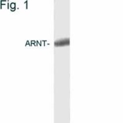
- Western Blot: ARNT/HIF-1 beta Antibody (2B10) [NB300-525] - Analysis of COS cell extract.

Supportive validation
- Submitted by
- Novus Biologicals (provider)
- Main image
- Experimental details
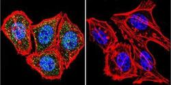
- Immunocytochemistry/Immunofluorescence: ARNT/HIF-1 beta Antibody (2B10) [NB300-525] - HIF-1 beta Antibody (2B10) [NB300-525] - HIF-1 beta (green), F-Actin staining with Phalloidin (red) and nuclei with DAPI (blue) is shown. Cells were grown on chamber slides and fixed with formaldehyde prior to staining. Cells were probed without (control) or with an antibody recognizing HIF-1 beta at a dilution of 1:20 over night at 4C, washed with PBS and incubated with a DyLight-488 conjugated.

- Submitted by
- Novus Biologicals (provider)
- Main image
- Experimental details
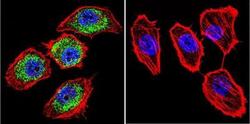
- Immunocytochemistry/Immunofluorescence: ARNT/HIF-1 beta Antibody (2B10) [NB300-525] - HIF-1 beta Antibody (2B10) [NB300-525] - HIF-1 beta (green), F-Actin staining with Phalloidin (red) and nuclei with DAPI (blue) is shown. Cells were grown on chamber slides and fixed with formaldehyde prior to staining. Cells were probed without (control) or with an antibody recognizing HIF-1 beta at a dilution of 1:100 over night at 4C, washed with PBS and incubated with a DyLight-488 conjugated.

- Submitted by
- Novus Biologicals (provider)
- Main image
- Experimental details
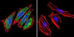
- Immunocytochemistry/Immunofluorescence: ARNT/HIF-1 beta Antibody (2B10) [NB300-525] - HIF-1 beta (green), F-Actin staining with Phalloidin (red) and nuclei with DAPI (blue) is shown. Cells were grown on chamber slides and fixed with formaldehyde prior to staining. Cells were probed without (control) or with an antibody recognizing HIF-1 beta at a dilution of 1:100 over night at 4C, washed with PBS and incubated with a DyLight-488 conjugated.

- Submitted by
- Novus Biologicals (provider)
- Main image
- Experimental details
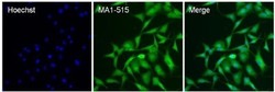
- Immunocytochemistry/Immunofluorescence: ARNT/HIF-1 beta Antibody (2B10) [NB300-525] - Analysis of HIF-1 beta (green) in NRK cells. The cells were fixed with 4% paraformaldehyde for 15 minutes, permeabilized with 0.1% Triton X-100 in PBS for 15 minutes, and blocked with 3% BSA in PBS for 30 minutes at room temperature. Cells were stained with a HIF-1 beta antibody at a dilution of 40ug/mL in staining buffer for 1 hour at room temperature, and then incubated with a Goat anti-Mouse IgG (H+L) Superclonal Secondary Antibody, Alexa Fluor 488 conjugate at a dilution of 1:1000 for 1 hour at room temperature (green). Nuclei (blue) were stained with Hoechst 33342 dye. Images were taken on a Thermo Scientific ToxInsight Instrument at 20X magnification.
