Explore
 Validate
 Learn
Learn Western blot
 Immunocytochemistry
Antibody data
- Antibody Data
- Antigen structure
- References [2]
- Comments [0]
- Validations
- Immunocytochemistry [2]
Submit
Validation data
Reference
Comment
Report error
- Product number
- GTX23533 - Provider product page

- Provider
- GeneTex
- Proper citation
- GeneTex Cat#GTX23533, RRID:AB_385097
- Product name
- PC2 antibody
- Antibody type
- Polyclonal
- Reactivity
- Human, Mouse
- Host
- Rabbit
Submitted references PTBP1 is required for glucose-stimulated cap-independent translation of insulin granule proteins and Coxsackieviruses in beta cells.
Wolfram syndrome 1 gene (WFS1) product localizes to secretory granules and determines granule acidification in pancreatic beta-cells.
Knoch KP, Nath-Sain S, Petzold A, Schneider H, Beck M, Wegbrod C, Sönmez A, Münster C, Friedrich A, Roivainen M, Solimena M
Molecular metabolism 2014 Aug;3(5):518-30
Molecular metabolism 2014 Aug;3(5):518-30
Wolfram syndrome 1 gene (WFS1) product localizes to secretory granules and determines granule acidification in pancreatic beta-cells.
Hatanaka M, Tanabe K, Yanai A, Ohta Y, Kondo M, Akiyama M, Shinoda K, Oka Y, Tanizawa Y
Human molecular genetics 2011 Apr 1;20(7):1274-84
Human molecular genetics 2011 Apr 1;20(7):1274-84
No comments: Submit comment
Supportive validation
- Submitted by
- GeneTex (provider)
- Main image
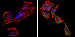
- Experimental details
- Immunofluorescent analysis of PC2 (green) showing staining in the cytoplasm of C2C12 cells. Formalin-fixed cells were permeabilized with 0.1% Triton X-100 in TBS for 5-10 minutes and blocked with 3% BSA-PBS for 30 minutes at room temperature. Cells were probed with a PC2 polyclonal antibody (GTX23533) in 3% BSA-PBS at a dilution of 1:100 and incubated overnight at 4 ?C in a humidified chamber. Cells were washed with PBST and incubated with a DyLight-conjugated secondary antibody in PBS at room temperature in the dark. F-actin (red) was stained with a flourescent red phalloidin and nuclei (blue) were stained with Hoechst or DAPI. Images were taken at a magnification of 60x.

- Submitted by
- GeneTex (provider)
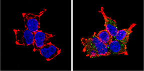
- Main image

- Experimental details
- Immunofluorescent analysis of PC2 (green) showing staining in the cytoplasm of HEK293 cells. Formalin-fixed cells were permeabilized with 0.1% Triton X-100 in TBS for 5-10 minutes and blocked with 3% BSA-PBS for 30 minutes at room temperature. Cells were probed with a PC2 polyclonal antibody (GTX23533) in 3% BSA-PBS at a dilution of 1:100 and incubated overnight at 4 ?C in a humidified chamber. Cells were washed with PBST and incubated with a DyLight-conjugated secondary antibody in PBS at room temperature in the dark. F-actin (red) was stained with a flourescent red phalloidin and nuclei (blue) were stained with Hoechst or DAPI. Images were taken at a magnification of 60x.