Explore
 Validate
 Learn
Learn Western blot
 Immunocytochemistry
Antibody data
- Antibody Data
- Antigen structure
- References [4]
- Comments [0]
- Validations
- Immunocytochemistry [3]
Submit
Validation data
Reference
Comment
Report error
- Product number
- PA1-058 - Provider product page

- Provider
- Invitrogen Antibodies
- Product name
- PCSK2 Polyclonal Antibody
- Antibody type
- Polyclonal
- Antigen
- Synthetic peptide
- Description
- PA1-058 detects prohormone convertase 2 (PC2) from human and mouse alpha pancreas cells. PA1-058 has been successfully used in ICC/IF and Western blot procedures. By Western blot, this antibody detects an ~65 kDa protein representing PC2 from alpha TC 6 cell extract. This antibody is not recommended for Neuro-2a cells in immunfluorescent applications. The PA1-058 immunogen is a synthetic peptide corresponding to residues E(622) L D E A V E R S L Q S I L R K N(638) of mouse PC2. This sequence is completely conserved between mouse and rat protein. PA1-058 synthetic peptide (PEP-138) is available for use in neutralization and control experiments.
- Reactivity
- Human, Mouse
- Host
- Rabbit
- Isotype
- IgG
- Vial size
- 100 μg
- Concentration
- 1 mg/mL
- Storage
- -20°C, Avoid Freeze/Thaw Cycles
Submitted references Revisiting Proinsulin Processing: Evidence That Human β-Cells Process Proinsulin With Prohormone Convertase (PC) 1/3 but Not PC2.
Chromogranin B regulates early-stage insulin granule trafficking from the Golgi in pancreatic islet β-cells.
Insulin-Deficient Mouse β-Cells Do Not Fully Mature but Can Be Remedied Through Insulin Replacement by Islet Transplantation.
Compensatory Islet Response to Insulin Resistance Revealed by Quantitative Proteomics.
Ramzy A, Asadi A, Kieffer TJ
Diabetes 2020 Jul;69(7):1451-1462
Diabetes 2020 Jul;69(7):1451-1462
Chromogranin B regulates early-stage insulin granule trafficking from the Golgi in pancreatic islet β-cells.
Bearrows SC, Bauchle CJ, Becker M, Haldeman JM, Swaminathan S, Stephens SB
Journal of cell science 2019 Jul 1;132(13)
Journal of cell science 2019 Jul 1;132(13)
Insulin-Deficient Mouse β-Cells Do Not Fully Mature but Can Be Remedied Through Insulin Replacement by Islet Transplantation.
Ramzy A, Mojibian M, Kieffer TJ
Endocrinology 2018 Jan 1;159(1):83-102
Endocrinology 2018 Jan 1;159(1):83-102
Compensatory Islet Response to Insulin Resistance Revealed by Quantitative Proteomics.
El Ouaamari A, Zhou JY, Liew CW, Shirakawa J, Dirice E, Gedeon N, Kahraman S, De Jesus DF, Bhatt S, Kim JS, Clauss TR, Camp DG 2nd, Smith RD, Qian WJ, Kulkarni RN
Journal of proteome research 2015 Aug 7;14(8):3111-3122
Journal of proteome research 2015 Aug 7;14(8):3111-3122
No comments: Submit comment
Supportive validation
- Submitted by
- Invitrogen Antibodies (provider)
- Main image
- Experimental details
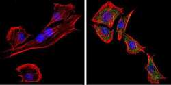
- Immunofluorescent analysis of PC2 (green) showing staining in the cytoplasm of C2C12 cells. Formalin-fixed cells were permeabilized with 0.1% Triton X-100 in TBS for 5-10 minutes and blocked with 3% BSA-PBS for 30 minutes at room temperature. Cells were probed with a PC2 polyclonal antibody (Product # PA1-058) in 3% BSA-PBS at a dilution of 1:100 and incubated overnight at 4 ºC in a humidified chamber. Cells were washed with PBST and incubated with a DyLight-conjugated secondary antibody in PBS at room temperature in the dark. F-actin (red) was stained with a fluorescent red phalloidin and nuclei (blue) were stained with Hoechst or DAPI. Images were taken at a magnification of 60x.
- Submitted by
- Invitrogen Antibodies (provider)
- Main image
- Experimental details
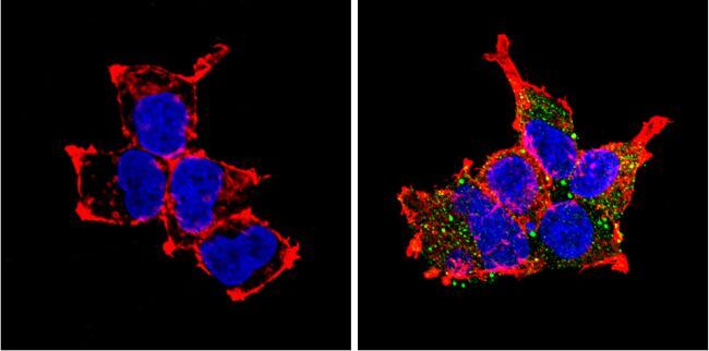
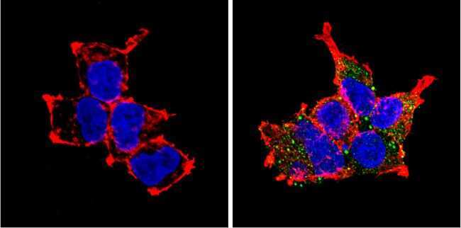
- Immunofluorescent analysis of PC2 (green) showing staining in the cytoplasm of HEK293 cells. Formalin-fixed cells were permeabilized with 0.1% Triton X-100 in TBS for 5-10 minutes and blocked with 3% BSA-PBS for 30 minutes at room temperature. Cells were probed with a PC2 polyclonal antibody (Product # PA1-058) in 3% BSA-PBS at a dilution of 1:100 and incubated overnight at 4 ºC in a humidified chamber. Cells were washed with PBST and incubated with a DyLight-conjugated secondary antibody in PBS at room temperature in the dark. F-actin (red) was stained with a fluorescent red phalloidin and nuclei (blue) were stained with Hoechst or DAPI. Images were taken at a magnification of 60x.
- Submitted by
- Invitrogen Antibodies (provider)
- Main image

- Experimental details
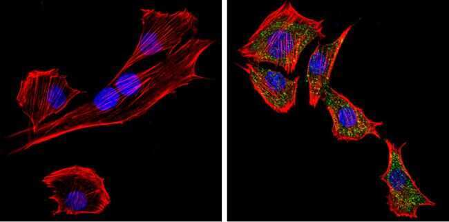
- Immunofluorescent analysis of PC2 (green) showing staining in the cytoplasm of HEK293 cells. Formalin-fixed cells were permeabilized with 0.1% Triton X-100 in TBS for 5-10 minutes and blocked with 3% BSA-PBS for 30 minutes at room temperature. Cells were probed with a PC2 polyclonal antibody (Product # PA1-058) in 3% BSA-PBS at a dilution of 1:100 and incubated overnight at 4 ºC in a humidified chamber. Cells were washed with PBST and incubated with a DyLight-conjugated secondary antibody in PBS at room temperature in the dark. F-actin (red) was stained with a fluorescent red phalloidin and nuclei (blue) were stained with Hoechst or DAPI. Images were taken at a magnification of 60x.