Explore
 Validate
 Learn
Learn Western blot
 Immunocytochemistry
Antibody data
- Antibody Data
- Antigen structure
- References [10]
- Comments [0]
- Validations
- Immunocytochemistry [2]
- Immunohistochemistry [4]
- Flow cytometry [1]
- Other assay [3]
Submit
Validation data
Reference
Comment
Report error
- Product number
- 42-0400 - Provider product page

- Provider
- Invitrogen Antibodies
- Product name
- SMAD7 Polyclonal Antibody
- Antibody type
- Polyclonal
- Antigen
- Synthetic peptide
- Reactivity
- Human, Mouse, Rat
- Host
- Rabbit
- Isotype
- IgG
- Vial size
- 100 μg
- Concentration
- 0.25 mg/mL
- Storage
- -20°C
Submitted references Deletion of Akt1 Promotes Kidney Fibrosis in a Murine Model of Unilateral Ureteral Obstruction.
Cancer-associated fibroblasts downregulate type I interferon receptor to stimulate intratumoral stromagenesis.
Role of sialidase Neu3 and ganglioside GM3 in cardiac fibroblasts activation.
HDAC inhibitors promote pancreatic stellate cell apoptosis and relieve pancreatic fibrosis by upregulating miR-15/16 in chronic pancreatitis.
Effects of tacrolimus on the TGF‑β1/SMAD signaling pathway in paraquat‑exposed rat alveolar type II epithelial cells.
Cryptotanshinone Protects Cartilage against Developing Osteoarthritis through the miR-106a-5p/GLIS3 Axis.
SMAD7 regulates proinflammatory and prolabor mediators in amnion and myometrium.
Cucurbitacin I Attenuates Cardiomyocyte Hypertrophy via Inhibition of Connective Tissue Growth Factor (CCN2) and TGF- β/Smads Signalings.
Neuropilin-2 Is upregulated in lung cancer cells during TGF-β1-induced epithelial-mesenchymal transition.
Kruppel-like factor KLF10 targets transforming growth factor-beta1 to regulate CD4(+)CD25(-) T cells and T regulatory cells.
Kim IY, Lee MY, Park MW, Seong EY, Lee DW, Lee SB, Bae SS, Kim SS, Song SH
BioMed research international 2020;2020:6143542
BioMed research international 2020;2020:6143542
Cancer-associated fibroblasts downregulate type I interferon receptor to stimulate intratumoral stromagenesis.
Cho C, Mukherjee R, Peck AR, Sun Y, McBrearty N, Katlinski KV, Gui J, Govindaraju PK, Puré E, Rui H, Fuchs SY
Oncogene 2020 Sep;39(38):6129-6137
Oncogene 2020 Sep;39(38):6129-6137
Role of sialidase Neu3 and ganglioside GM3 in cardiac fibroblasts activation.
Ghiroldi A, Piccoli M, Creo P, Cirillo F, Rota P, D'Imperio S, Ciconte G, Monasky MM, Micaglio E, Garatti A, Aureli M, Carsana EV, Menicanti L, Pappone C, Anastasia L
The Biochemical journal 2020 Sep 18;477(17):3401-3415
The Biochemical journal 2020 Sep 18;477(17):3401-3415
HDAC inhibitors promote pancreatic stellate cell apoptosis and relieve pancreatic fibrosis by upregulating miR-15/16 in chronic pancreatitis.
Ji T, Feng W, Zhang X, Zang K, Zhu X, Shang F
Human cell 2020 Oct;33(4):1006-1016
Human cell 2020 Oct;33(4):1006-1016
Effects of tacrolimus on the TGF‑β1/SMAD signaling pathway in paraquat‑exposed rat alveolar type II epithelial cells.
Ren Y, Jian X, Zhang Z, Ning Q, Kan B, Kong L
Molecular medicine reports 2020 Nov;22(5):3687-3694
Molecular medicine reports 2020 Nov;22(5):3687-3694
Cryptotanshinone Protects Cartilage against Developing Osteoarthritis through the miR-106a-5p/GLIS3 Axis.
Ji Q, Qi D, Xu X, Xu Y, Goodman SB, Kang L, Song Q, Fan Z, Maloney WJ, Wang Y
Molecular therapy. Nucleic acids 2018 Jun 1;11:170-179
Molecular therapy. Nucleic acids 2018 Jun 1;11:170-179
SMAD7 regulates proinflammatory and prolabor mediators in amnion and myometrium.
Lim R, Barker G, Lappas M
Biology of reproduction 2017 Aug 1;97(2):288-301
Biology of reproduction 2017 Aug 1;97(2):288-301
Cucurbitacin I Attenuates Cardiomyocyte Hypertrophy via Inhibition of Connective Tissue Growth Factor (CCN2) and TGF- β/Smads Signalings.
Jeong MH, Kim SJ, Kang H, Park KW, Park WJ, Yang SY, Yang DK
PloS one 2015;10(8):e0136236
PloS one 2015;10(8):e0136236
Neuropilin-2 Is upregulated in lung cancer cells during TGF-β1-induced epithelial-mesenchymal transition.
Nasarre P, Gemmill RM, Potiron VA, Roche J, Lu X, Barón AE, Korch C, Garrett-Mayer E, Lagana A, Howe PH, Drabkin HA
Cancer research 2013 Dec 1;73(23):7111-21
Cancer research 2013 Dec 1;73(23):7111-21
Kruppel-like factor KLF10 targets transforming growth factor-beta1 to regulate CD4(+)CD25(-) T cells and T regulatory cells.
Cao Z, Wara AK, Icli B, Sun X, Packard RR, Esen F, Stapleton CJ, Subramaniam M, Kretschmer K, Apostolou I, von Boehmer H, Hansson GK, Spelsberg TC, Libby P, Feinberg MW
The Journal of biological chemistry 2009 Sep 11;284(37):24914-24
The Journal of biological chemistry 2009 Sep 11;284(37):24914-24
No comments: Submit comment
Supportive validation
- Submitted by
- Invitrogen Antibodies (provider)
- Main image

- Experimental details
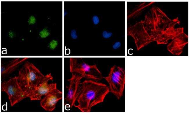
- Immunofluorescence analysis of SMAD7 was done on 70% confluent log phase HeLa cells. The cells were fixed with 4% paraformaldehyde for 15 minutes, permeabilized with 0.25% Triton™ X-100 for 10 minutes, and blocked with 5% BSA for 1 hour at room temperature. The cells were labeled with SMAD7 Rabbit Polyclonal Antibody (Product # 42-0400) at 1 µg/mL in 1% BSA and incubated for 3 hours at room temperature and then labeled with Goat anti-Rabbit IgG (H+L) Superclonal™ Secondary Antibody, Alexa Fluor® 488 conjugate (Product # A27034) at a dilution of 1:2000 for 45 minutes at room temperature (Panel a: green). Nuclei (Panel b: blue) were stained with SlowFade® Gold Antifade Mountant with DAPI (Product # S36938). F-actin (Panel c: red) was stained with Alexa Fluor® 555 Rhodamine Phalloidin (Product # R415, 1:300). Panel d is a merged image showing Nuclear localization. Panel e is a no primary antibody control. The images were captured at 60X magnification.
- Submitted by
- Invitrogen Antibodies (provider)
- Main image
- Experimental details
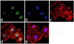
- Immunofluorescence analysis of SMAD7 was done on 70% confluent log phase HeLa cells. The cells were fixed with 4% paraformaldehyde for 15 minutes, permeabilized with 0.25% Triton™ X-100 for 10 minutes, and blocked with 5% BSA for 1 hour at room temperature. The cells were labeled with SMAD7 Rabbit Polyclonal Antibody (Product # 42-0400) at 1 µg/mL in 1% BSA and incubated for 3 hours at room temperature and then labeled with Goat anti-Rabbit IgG (Heavy Chain) Superclonal™ Secondary Antibody, Alexa Fluor® 488 conjugate (Product # A27034) at a dilution of 1:2000 for 45 minutes at room temperature (Panel a: green). Nuclei (Panel b: blue) were stained with SlowFade® Gold Antifade Mountant with DAPI (Product # S36938). F-actin (Panel c: red) was stained with Alexa Fluor® 555 Rhodamine Phalloidin (Product # R415, 1:300). Panel d is a merged image showing Nuclear localization. Panel e is a no primary antibody control. The images were captured at 60X magnification.

Supportive validation
- Submitted by
- Invitrogen Antibodies (provider)
- Main image
- Experimental details
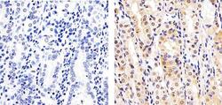
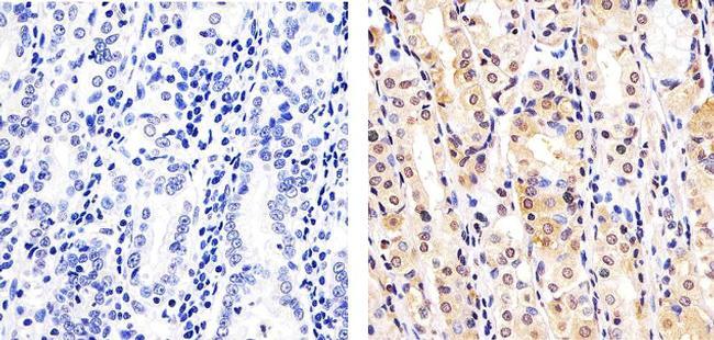
- Immunohistochemistry analysis of SMAD7 showing staining in the cytoplasm and nucleus of paraffin-embedded human stomach tissue (right) compared to a negative control without primary antibody (left). To expose target proteins, antigen retrieval was performed using 10mM sodium citrate (pH 6.0), microwaved for 8-15 min. Following antigen retrieval, tissues were blocked in 3% H2O2-methanol for 15 min at room temperature, washed with ddH2O and PBS, and then probed with a SMAD7 Rabbit Polyclonal Antibody (Product # 42-0400) diluted in 3% BSA-PBS at a dilution of 1:20 overnight at 4ºC in a humidified chamber. Tissues were washed extensively in PBST and detection was performed using an HRP-conjugated secondary antibody followed by colorimetric detection using a DAB kit. Tissues were counterstained with hematoxylin and dehydrated with ethanol and xylene to prep for mounting.
- Submitted by
- Invitrogen Antibodies (provider)
- Main image

- Experimental details
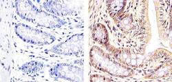
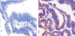
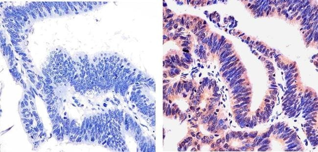
- Immunohistochemistry analysis of SMAD7 showing staining in the cytoplasm and nucleus of paraffin-embedded human colon carcinoma (right) compared to a negative control without primary antibody (left). To expose target proteins, antigen retrieval was performed using 10mM sodium citrate (pH 6.0), microwaved for 8-15 min. Following antigen retrieval, tissues were blocked in 3% H2O2-methanol for 15 min at room temperature, washed with ddH2O and PBS, and then probed with a SMAD7 Rabbit Polyclonal Antibody (Product # 42-0400) diluted in 3% BSA-PBS at a dilution of 1:20 overnight at 4°C in a humidified chamber. Tissues were washed extensively in PBST and detection was performed using an HRP-conjugated secondary antibody followed by colorimetric detection using a DAB kit. Tissues were counterstained with hematoxylin and dehydrated with ethanol and xylene to prep for mounting.

- Submitted by
- Invitrogen Antibodies (provider)
- Main image
- Experimental details
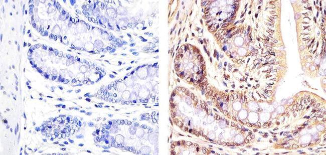
- Immunohistochemistry analysis of SMAD7 showing staining in the cytoplasm and nucleus of paraffin-embedded rat rectum tissue (right) compared to a negative control without primary antibody (left). To expose target proteins, antigen retrieval was performed using 10mM sodium citrate (pH 6.0), microwaved for 8-15 min. Following antigen retrieval, tissues were blocked in 3% H2O2-methanol for 15 min at room temperature, washed with ddH2O and PBS, and then probed with a SMAD7 Rabbit Polyclonal Antibody (Product # 42-0400) diluted in 3% BSA-PBS at a dilution of 1:20 overnight at 4ºC in a humidified chamber. Tissues were washed extensively in PBST and detection was performed using an HRP-conjugated secondary antibody followed by colorimetric detection using a DAB kit. Tissues were counterstained with hematoxylin and dehydrated with ethanol and xylene to prep for mounting.
- Submitted by
- Invitrogen Antibodies (provider)
- Main image
- Experimental details
- Immunohistochemistry analysis of SMAD7 showing staining in the cytoplasm and nucleus of paraffin-embedded human colon carcinoma (right) compared to a negative control without primary antibody (left). To expose target proteins, antigen retrieval was performed using 10mM sodium citrate (pH 6.0), microwaved for 8-15 min. Following antigen retrieval, tissues were blocked in 3% H2O2-methanol for 15 min at room temperature, washed with ddH2O and PBS, and then probed with a SMAD7 Rabbit Polyclonal Antibody (Product # 42-0400) diluted in 3% BSA-PBS at a dilution of 1:20 overnight at 4°C in a humidified chamber. Tissues were washed extensively in PBST and detection was performed using an HRP-conjugated secondary antibody followed by colorimetric detection using a DAB kit. Tissues were counterstained with hematoxylin and dehydrated with ethanol and xylene to prep for mounting.
Supportive validation
- Submitted by
- Invitrogen Antibodies (provider)
- Main image
- Experimental details
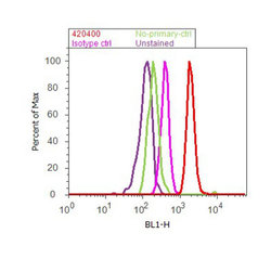
- Flow cytometry analysis of SMAD7 was done on Jurkat cells treated with TNF alpha (50ng/mL, 20 minutes). Cells were fixed with 70% ethanol for 10 minutes, permeabilized with 0.25% Triton™ X-100 for 20 minutes, and blocked with 5% BSA for 30 minutes at room temperature. Cells were labeled with SMAD7 Rabbit Polyclonal Antibody (420400, red histogram) or with rabbit isotype control (pink histogram) at 3-5 ug/million cells in 2.5% BSA. After incubation at room temperature for 2 hours, the cells were labeled with Alexa Fluor® 488 Goat Anti-Rabbit Secondary Antibody (A11008) at a dilution of 1:400 for 30 minutes at room temperature. The representative 10,000 cells were acquired and analyzed for each sample using an Attune® Acoustic Focusing Cytometer. The purple histogram represents unstained control cells and the green histogram represents no-primary-antibody control.
Supportive validation
- Submitted by
- Invitrogen Antibodies (provider)
- Main image
- Experimental details
- NULL
- Submitted by
- Invitrogen Antibodies (provider)
- Main image
- Experimental details
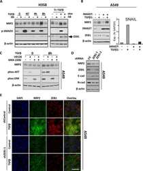
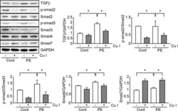
- Fig 4 Cucurbitacin I blocks the TGF-beta/Smad signaling pathway in hypertrophic cardiomyocytes. Cell extracts (50 mug) were used for Western blot analysis of TGF-beta and Smad (Smad2, 3, and 7) protein expression levels. The expression levels of TGF-beta, Smad2, 3, and 7, and the phosphorylated forms of Smad2 and 3 (p-Smad2 and p-Smad3) were estimated by measuring band densities with NIH Image J software. GAPDH was used as a loading control. And Western blot analysis was performed in triplicate with three independent samples. Data are expressed as fold changes +- S.D. vs. control group. Significance was measured via a two-way ANOVA. * P < 0.05. Cont, control; Cu I, Cucurbitacin I.

- Submitted by
- Invitrogen Antibodies (provider)
- Main image
- Experimental details
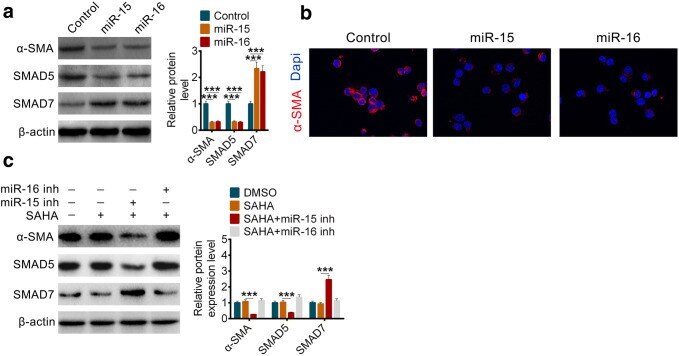
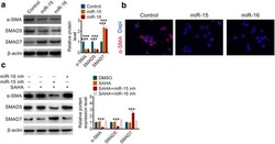
- Fig. 5 HDAC inhibition disrupts TGF-beta/Smad Signaling by miR-15 and miR-16 to protect against pancreatic fibrosis. a The protein levels and the related quantification of alpha-SMA, SMAD5 and SMAD7 in PSCs after overexpression of miR-15 or miR-16. b Immunofluorescence staining of alpha-SMA in PSCs after overexpression of miR-15 or miR-16. c The protein levels and the related quantification of alpha-SMA, SMAD5 and SMAD7 in PSCs after transfected with miR-15 inhibitor or miR-16 inhibitor with or without SAHA. All data are presented as the mean +- S.D. ( n = 3). *** P < 0.001, compared with control. Each assay was performed in triplicate