Explore
 Validate
 Learn
Learn Immunocytochemistry
Antibody data
- Antibody Data
- Antigen structure
- References [4]
- Comments [0]
- Validations
- Immunocytochemistry [1]
- Immunohistochemistry [1]
Submit
Validation data
Reference
Comment
Report error
- Product number
- HPA005922 - Provider product page

- Provider
- Atlas Antibodies
- Proper citation
- Atlas Antibodies Cat#HPA005922, RRID:AB_1078860
- Product name
- Anti-FHL2
- Antibody type
- Polyclonal
- Description
- Polyclonal Antibody against Human FHL2, Gene description: four and a half LIM domains 2, Alternative Gene Names: DRAL, SLIM3, Validated applications: ICC, IHC, Uniprot ID: Q14192, Storage: Store at +4°C for short term storage. Long time storage is recommended at -20°C.
- Reactivity
- Human
- Host
- Rabbit
- Conjugate
- Unconjugated
- Isotype
- IgG
- Vial size
- 100 µl
- Concentration
- 0.1 mg/ml
- Storage
- Store at +4°C for short term storage. Long time storage is recommended at -20°C.
- Handling
- The antibody solution should be gently mixed before use.
Submitted references fhl2b mediates extraocular muscle protection in zebrafish models of muscular dystrophies and its ectopic expression ameliorates affected body muscles.
Immunofluorescence and fluorescent-protein tagging show high correlation for protein localization in mammalian cells
Systematic validation of antibody binding and protein subcellular localization using siRNA and confocal microscopy
The interactome of LIM domain proteins: The contributions of LIM domain proteins to heart failure and heart development
Dennhag N, Kahsay A, Nissen I, Nord H, Chermenina M, Liu J, Arner A, Liu JX, Backman LJ, Remeseiro S, von Hofsten J, Pedrosa Domellöf F
Nature communications 2024 Mar 2;15(1):1950
Nature communications 2024 Mar 2;15(1):1950
Immunofluorescence and fluorescent-protein tagging show high correlation for protein localization in mammalian cells
Stadler C, Rexhepaj E, Singan V, Murphy R, Pepperkok R, Uhlén M, Simpson J, Lundberg E
Nature Methods 2013;10(4):315-323
Nature Methods 2013;10(4):315-323
Systematic validation of antibody binding and protein subcellular localization using siRNA and confocal microscopy
Stadler C, Hjelmare M, Neumann B, Jonasson K, Pepperkok R, Uhlén M, Lundberg E
Journal of Proteomics 2012;75(7):2236-2251
Journal of Proteomics 2012;75(7):2236-2251
The interactome of LIM domain proteins: The contributions of LIM domain proteins to heart failure and heart development
Li A, Ponten F, dos Remedios C
PROTEOMICS 2012;12(2):203-225
PROTEOMICS 2012;12(2):203-225
No comments: Submit comment
Supportive validation
- Submitted by
- Atlas Antibodies (provider)
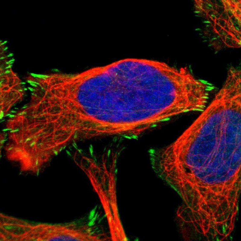
- Main image
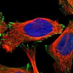
- Experimental details
- Immunofluorescent staining of human cell line U-2 OS shows localization to actin filaments & focal adhesion sites.
- Sample type
- Human
Supportive validation
- Submitted by
- Atlas Antibodies (provider)
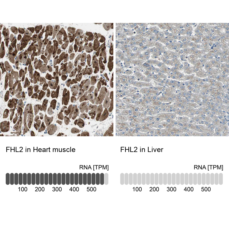
- Enhanced method
- Orthogonal validation
- Main image
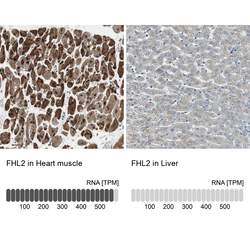
- Experimental details
- Immunohistochemistry analysis in human heart muscle and liver tissues using HPA005922 antibody. Corresponding FHL2 RNA-seq data are presented for the same tissues.
- Sample type
- Human
- Protocol
- Protocol