 Explore
 Validate
 Learn
Learn Western blot
 Immunocytochemistry
Antibody data
- Antibody Data
- Antigen structure
- References [1]
- Comments [0]
- Validations
- Immunocytochemistry [1]
- Immunohistochemistry [6]
- Other assay [2]
Submit
Validation data
Reference
Comment
Report error
- Product number
- PA5-64182 - Provider product page

- Provider
- Invitrogen Antibodies
- Product name
- LRP2 Polyclonal Antibody
- Antibody type
- Polyclonal
- Antigen
- Recombinant protein fragment
- Description
- Immunogen sequence: NNDRIYWSDF KEDVIETIKY DGTDRRVIAK EAMNPYSLDI FEDQLYWISK EKGEVWKQNK FGQGKKEKTL VVNPWLTQVR IFHQLRYNKS VP Highest antigen sequence identity to the following orthologs: Mouse - 82%, Rat - 79%.
- Reactivity
- Human
- Host
- Rabbit
- Isotype
- IgG
- Vial size
- 100 μL
- Concentration
- 0.1 mg/mL
- Storage
- Store at 4°C short term. For long term storage, store at -20°C, avoiding freeze/thaw cycles.
Submitted references Complete remission of nephrotic syndrome in a young woman with anti-LRP2 nephropathy after immunosuppressive therapy.
Zhu X, Tu L, Liu S, You H, Xue J, Hao C
BMC nephrology 2020 Aug 24;21(1):364
BMC nephrology 2020 Aug 24;21(1):364
No comments: Submit comment
Supportive validation
- Submitted by
- Invitrogen Antibodies (provider)
- Main image

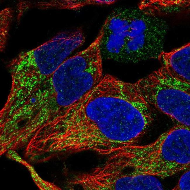
- Experimental details
- Immunofluorescent staining of LRP2 in human cell line HEK 293 shows positivity in mitochondria. Samples were probed using a LRP2 Polyclonal Antibody (Product # PA5-64182).
Supportive validation
- Submitted by
- Invitrogen Antibodies (provider)
- Main image

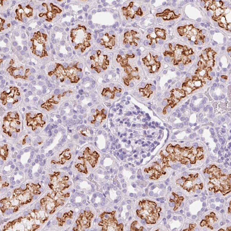
- Experimental details
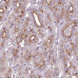
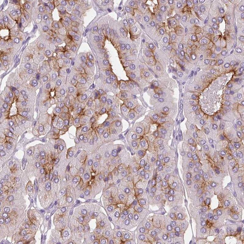
- Immunohistochemical analysis of LRP2 in human kidney using LRP2 Polyclonal Antibody (Product # PA5-64182) shows strong membranous positivity in cells in tubules.
- Submitted by
- Invitrogen Antibodies (provider)
- Main image
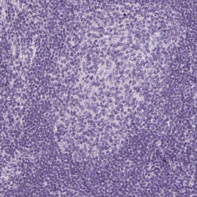
- Experimental details
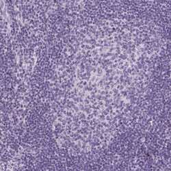
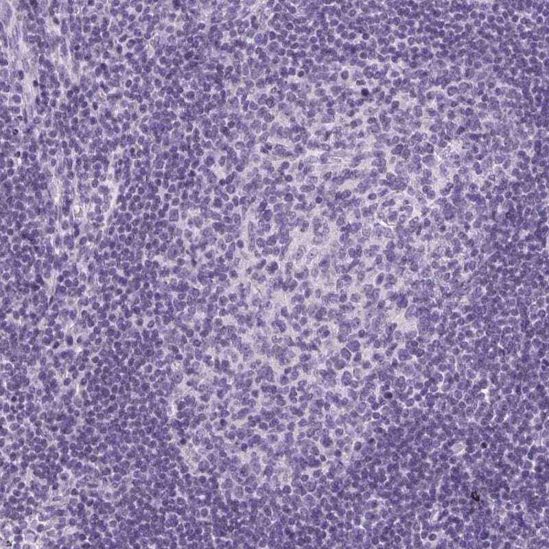
- Immunohistochemical analysis of LRP2 in human lymph node using LRP2 Polyclonal Antibody (Product # PA5-64182) shows no positivity in germinal center cells as expected.
- Submitted by
- Invitrogen Antibodies (provider)
- Main image
- Experimental details
- Immunohistochemical analysis of LRP2 in human parathyroid gland using LRP2 Polyclonal Antibody (Product # PA5-64182) shows strong membranous positivity in glandular cells.
- Submitted by
- Invitrogen Antibodies (provider)
- Main image
- Experimental details
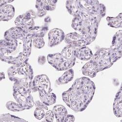
- Immunohistochemical analysis of LRP2 in human placenta using LRP2 Polyclonal Antibody (Product # PA5-64182) shows no positivity in trophoblastic cells as expected.

- Submitted by
- Invitrogen Antibodies (provider)
- Main image
- Experimental details
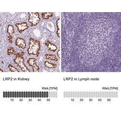
- Immunohistochemical staining of LRP2 in human kidney and lymph node tissues using LRP2 Polyclonal Antibody (Product # PA5-64182). Corresponding LRP2 RNA-seq data are presented for the same tissues.

- Submitted by
- Invitrogen Antibodies (provider)
- Main image
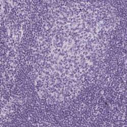
- Experimental details
- Immunohistochemical staining of LRP2 in human lymph node using LRP2 Polyclonal Antibody (Product # PA5-64182) shows low expression as expected.
Supportive validation
- Submitted by
- Invitrogen Antibodies (provider)
- Main image

- Experimental details
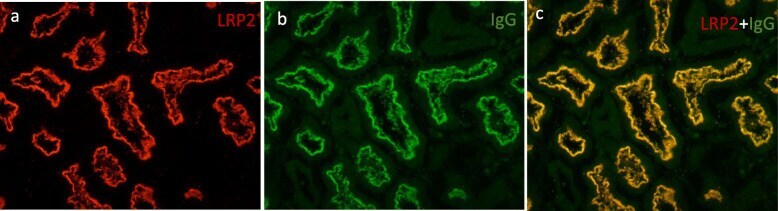
- Fig. 2 Colocalization of LRP2 and IgG on the patient's renal biopsy sample. a - c Immunofluorescence experiments showed positive staining along the apical membrane of the proximal tubules for: a LRP2 using a rabbit polyclonal antibody and b IgG. c Strong colocalization of LRP2 and IgG along the apical membrane of the proximal tubules
- Submitted by
- Invitrogen Antibodies (provider)
- Main image

- Experimental details
- Fig. 4 Immunofluorescence performed on cryosections of normal human kidney tissue. a Staining for LRP2 using a rabbit antibody along the apical membrane of the proximal tubular epithelium. b Indirect immunofluorescence of serum from a patient with anti-LRP2 nephropathy on a normal human kidney. c Strong colocalization of LRP2 and serum antibodies
