 Explore
 Validate
 Learn
Learn Western blot
 Immunocytochemistry
Antibody data
- Antibody Data
- Antigen structure
- References [0]
- Comments [0]
- Validations
- Immunocytochemistry [5]
- Immunoprecipitation [1]
- Immunohistochemistry [3]
- Other assay [1]
Submit
Validation data
Reference
Comment
Report error
- Product number
- PA5-22102 - Provider product page

- Provider
- Invitrogen Antibodies
- Product name
- GLDC Polyclonal Antibody
- Antibody type
- Polyclonal
- Antigen
- Recombinant full-length protein
- Description
- Recommended positive controls: HePG2. Predicted reactivity: Mouse (91%), Rat (84%), Xenopus laevis (86%), Chicken (85%), Bovine (92%). Store product as a concentrated solution. Centrifuge briefly prior to opening the vial.
- Reactivity
- Human, Mouse, Rat
- Host
- Rabbit
- Isotype
- IgG
- Vial size
- 100 μL
- Concentration
- 0.46 mg/mL
- Storage
- Store at 4°C short term. For long term storage, store at -20°C, avoiding freeze/thaw cycles.
No comments: Submit comment
Supportive validation
- Submitted by
- Invitrogen Antibodies (provider)
- Main image
- Experimental details
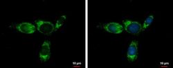
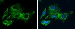
- Immunofluorescent analysis of glycine dehydrogenase (decarboxylating) precursor showing staining in the mitochondria of HepG2 cells. HepG2 cells were fixed in ice-cold MeOH for 5 min and stained using a glycine dehydrogenase (decarboxylating) precursor polyclonal antibody (Product # PA5-22102) diluted at 1:500. Blue: Hoechst 33342 staining.
- Submitted by
- Invitrogen Antibodies (provider)
- Main image

- Experimental details
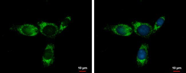
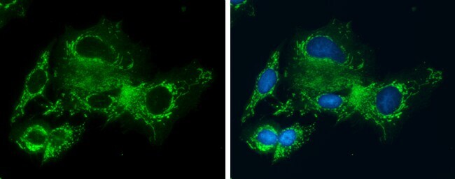
- Immunocytochemistry-Immunofluorescence analysis of GLDC was performed in Hep G2 cells fixed in ice-cold MeOH for 5 min. Green: GLDC Polyclonal Antibody (Product # PA5-22102) diluted at 1:400. Blue: Hoechst 33342 staining.

- Submitted by
- Invitrogen Antibodies (provider)
- Main image
- Experimental details
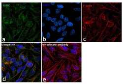
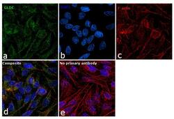
- Immunofluorescence analysis of GLDC was performed using 70% confluent log phase HeLa cells. The cells were fixed with ice-cold acetone for 5 minutes and blocked with 1% BSA for 1 hour at room temperature. The cells were labeled with GLDC Polyclonal Antibody (Product # PA5-22102) at 1:100 dilution in 0.1% BSA, incubated at 4 degree celsius overnight and then labeled with Goat anti-Rabbit IgG (H+L) Superclonal™ Secondary Antibody, Alexa Fluor® 488 conjugate (Product # A27034) at a dilution of 1:2000 for 45 minutes at room temperature (Panel a: green).Nuclei (Panel b: blue) were stained with SlowFade® Gold Antifade Mountant with DAPI (Product # S36938). F-actin (Panel c: red) was stained with Rhodamine Phalloidin (Product # R415, 1:300). Panel d represents the merged image showing mitochondrial and nuclear localization. Panel e represents control cells with no primary antibody to assess background. The images were captured at 60X magnification.
- Submitted by
- Invitrogen Antibodies (provider)
- Main image
- Experimental details
- Immunocytochemistry-Immunofluorescence analysis of GLDC was performed in Hep G2 cells fixed in ice-cold MeOH for 5 min. Green: GLDC Polyclonal Antibody (Product # PA5-22102) diluted at 1:400. Blue: Hoechst 33342 staining.
- Submitted by
- Invitrogen Antibodies (provider)
- Main image
- Experimental details
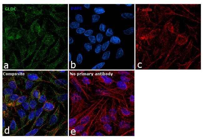
- Immunofluorescence analysis of GLDC was performed using 70% confluent log phase HeLa cells. The cells were fixed with ice-cold acetone for 5 minutes and blocked with 1% BSA for 1 hour at room temperature. The cells were labeled with GLDC Polyclonal Antibody (Product # PA5-22102) at 1:100 dilution in 0.1% BSA, incubated at 4 degree celsius overnight and then labeled with Goat anti-Rabbit IgG (Heavy Chain) Superclonal™ Secondary Antibody, Alexa Fluor® 488 conjugate (Product # A27034) at a dilution of 1:2000 for 45 minutes at room temperature (Panel a: green).Nuclei (Panel b: blue) were stained with SlowFade® Gold Antifade Mountant with DAPI (Product # S36938). F-actin (Panel c: red) was stained with Rhodamine Phalloidin (Product # R415, 1:300). Panel d represents the merged image showing mitochondrial and nuclear localization. Panel e represents control cells with no primary antibody to assess background. The images were captured at 60X magnification.

Supportive validation
- Submitted by
- Invitrogen Antibodies (provider)
- Main image
- Experimental details
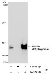
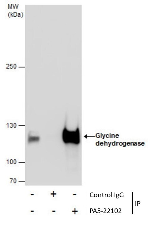
- Immunoprecipitation of Glycine dehydrogenase was performed in HepG2 whole cell extracts using 5 µg of GLDC Polyclonal Antibody (Product # PA5-22102). Samples were transferred to a membrane and probed with GLDC Polyclonal Antibody as a primary antibody and an HRP-conjugated anti-Rabbit IgG was used as a secondary antibody.
Supportive validation
- Submitted by
- Invitrogen Antibodies (provider)
- Main image
- Experimental details
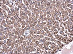
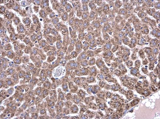
- Immunohistochemistry (Paraffin) analysis of GLDC was performed in paraffin-embedded mouse liver tissue using GLDC Polyclonal Antibody (Product # PA5-22102) at a dilution of 1:500.
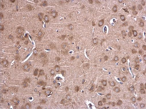
- Submitted by
- Invitrogen Antibodies (provider)
- Main image

- Experimental details
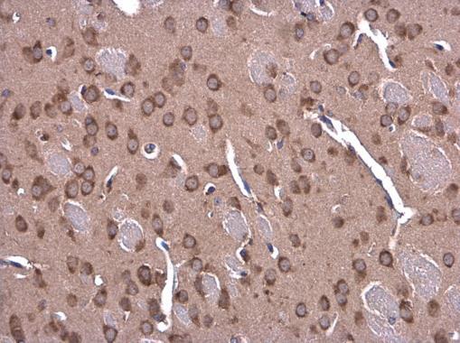
- Immunohistochemistry (Paraffin) analysis of GLDC was performed in paraffin-embedded mouse brain tissue using GLDC Polyclonal Antibody (Product # PA5-22102) at a dilution of 1:500.
- Submitted by
- Invitrogen Antibodies (provider)
- Main image
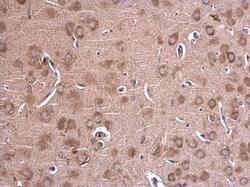
- Experimental details
- Immunohistochemistry (Paraffin) analysis of GLDC was performed in paraffin-embedded rat brain tissue using GLDC Polyclonal Antibody (Product # PA5-22102) at a dilution of 1:500.
Supportive validation
- Submitted by
- Invitrogen Antibodies (provider)
- Main image

- Experimental details
- Immunoprecipitation of Glycine dehydrogenase was performed in HepG2 whole cell extracts using 5 µg of GLDC Polyclonal Antibody (Product # PA5-22102). Samples were transferred to a membrane and probed with GLDC Polyclonal Antibody as a primary antibody and an HRP-conjugated anti-Rabbit IgG was used as a secondary antibody.
