Explore
 Validate
 Learn
Learn Western blot
 Immunocytochemistry
Antibody data
- Antibody Data
- Antigen structure
- References [11]
- Comments [0]
- Validations
- Western blot [1]
- Immunohistochemistry [3]
- Flow cytometry [8]
Submit
Validation data
Reference
Comment
Report error
- Product number
- NBP1-39474 - Provider product page

- Provider
- Novus Biologicals
- Proper citation
- Novus Cat#NBP1-39474, RRID:AB_2226520
- Product name
- Mouse Monoclonal Adenosine A2aR Antibody
- Antibody type
- Monoclonal
- Description
- Protein G purified.
- Reactivity
- Human, Mouse, Rat, Canine, Guinea Pig, Porcine, Rabbit, Sheep
- Host
- Mouse
- Antigen sequence
Epitope mapped to SQPLPGER in the t
hird intracellular loop (PMID 98221
47).- Isotype
- IgG
- Vial size
- 0.1 ml
- Concentration
- 1.0 mg/ml
- Storage
- Store at 4C short term. Aliquot and store at -20C long term. Avoid freeze-thaw cycles.
Submitted references Role of adenosine signaling in coordinating cardiomyocyte function and coronary vascular growth in chronic fetal anemia.
CD150high Bone Marrow Tregs Maintain Hematopoietic Stem Cell Quiescence and Immune Privilege via Adenosine.
A defect in KCa3.1 channel activity limits the ability of CD8+ T cells from cancer patients to infiltrate an adenosine-rich microenvironment.
A Novel Antagonist of the Immune Checkpoint Protein Adenosine A2a Receptor Restores Tumor-Infiltrating Lymphocyte Activity in the Context of the Tumor Microenvironment.
Altered expression of connexin 43 and related molecular partners in a pig model of left ventricular dysfunction with and without dipyrydamole therapy.
Melanoma Induces, and Adenosine Suppresses, CXCR3-Cognate Chemokine Production and T-cell Infiltration of Lungs Bearing Metastatic-like Disease.
Mechanisms involved in the adenosine-induced vasorelaxation to the pig prostatic small arteries.
Cross-regulation of carbon monoxide and the adenosine A2a receptor in macrophages.
Th1 cytokines regulate adenosine receptors and their downstream signaling elements in human microvascular endothelial cells.
Inflammatory cytokines regulate function and expression of adenosine A(2A) receptors in human monocytic THP-1 cells.
Immunohistochemical localization of adenosine A2A receptors in the rat central nervous system.
Davis L, Musso J, Soman D, Louey S, Nelson JW, Jonker SS
American journal of physiology. Regulatory, integrative and comparative physiology 2018 Sep 1;315(3):R500-R508
American journal of physiology. Regulatory, integrative and comparative physiology 2018 Sep 1;315(3):R500-R508
CD150high Bone Marrow Tregs Maintain Hematopoietic Stem Cell Quiescence and Immune Privilege via Adenosine.
Hirata Y, Furuhashi K, Ishii H, Li HW, Pinho S, Ding L, Robson SC, Frenette PS, Fujisaki J
Cell stem cell 2018 Mar 1;22(3):445-453.e5
Cell stem cell 2018 Mar 1;22(3):445-453.e5
A defect in KCa3.1 channel activity limits the ability of CD8+ T cells from cancer patients to infiltrate an adenosine-rich microenvironment.
Chimote AA, Balajthy A, Arnold MJ, Newton HS, Hajdu P, Qualtieri J, Wise-Draper T, Conforti L
Science signaling 2018 Apr 24;11(527)
Science signaling 2018 Apr 24;11(527)
A Novel Antagonist of the Immune Checkpoint Protein Adenosine A2a Receptor Restores Tumor-Infiltrating Lymphocyte Activity in the Context of the Tumor Microenvironment.
Mediavilla-Varela M, Castro J, Chiappori A, Noyes D, Hernandez DC, Allard B, Stagg J, Antonia SJ
Neoplasia (New York, N.Y.) 2017 Jul;19(7):530-536
Neoplasia (New York, N.Y.) 2017 Jul;19(7):530-536
Altered expression of connexin 43 and related molecular partners in a pig model of left ventricular dysfunction with and without dipyrydamole therapy.
Del Ry S, Moscato S, Bianchi F, Morales MA, Dolfi A, Burchielli S, Cabiati M, Mattii L
Pharmacological research 2015 May-Jun;95-96:92-101
Pharmacological research 2015 May-Jun;95-96:92-101
Melanoma Induces, and Adenosine Suppresses, CXCR3-Cognate Chemokine Production and T-cell Infiltration of Lungs Bearing Metastatic-like Disease.
Clancy-Thompson E, Perekslis TJ, Croteau W, Alexander MP, Chabanet TB, Turk MJ, Huang YH, Mullins DW
Cancer immunology research 2015 Aug;3(8):956-67
Cancer immunology research 2015 Aug;3(8):956-67
Mechanisms involved in the adenosine-induced vasorelaxation to the pig prostatic small arteries.
Ribeiro AS, Fernandes VS, Orensanz LM, Martínez MP, Recio P, Martínez-Sáenz A, Climent B, Arteaga JL, García-Sacristán A, Prieto D, Hernández M
Purinergic signalling 2011 Dec;7(4):413-25
Purinergic signalling 2011 Dec;7(4):413-25
Cross-regulation of carbon monoxide and the adenosine A2a receptor in macrophages.
Haschemi A, Wagner O, Marculescu R, Wegiel B, Robson SC, Gagliani N, Gallo D, Chen JF, Bach FH, Otterbein LE
Journal of immunology (Baltimore, Md. : 1950) 2007 May 1;178(9):5921-9
Journal of immunology (Baltimore, Md. : 1950) 2007 May 1;178(9):5921-9
Th1 cytokines regulate adenosine receptors and their downstream signaling elements in human microvascular endothelial cells.
Nguyen DK, Montesinos MC, Williams AJ, Kelly M, Cronstein BN
Journal of immunology (Baltimore, Md. : 1950) 2003 Oct 15;171(8):3991-8
Journal of immunology (Baltimore, Md. : 1950) 2003 Oct 15;171(8):3991-8
Inflammatory cytokines regulate function and expression of adenosine A(2A) receptors in human monocytic THP-1 cells.
Khoa ND, Montesinos MC, Reiss AB, Delano D, Awadallah N, Cronstein BN
Journal of immunology (Baltimore, Md. : 1950) 2001 Oct 1;167(7):4026-32
Journal of immunology (Baltimore, Md. : 1950) 2001 Oct 1;167(7):4026-32
Immunohistochemical localization of adenosine A2A receptors in the rat central nervous system.
Rosin DL, Robeva A, Woodard RL, Guyenet PG, Linden J
The Journal of comparative neurology 1998 Nov 16;401(2):163-86
The Journal of comparative neurology 1998 Nov 16;401(2):163-86
No comments: Submit comment
Supportive validation
- Submitted by
- Novus Biologicals (provider)
- Main image

- Experimental details
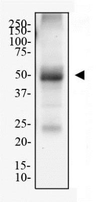
- Western Blot: Adenosine A2aR Antibody (7F6-G5-A2) [NBP1-39474] - Total protein from mouse brain was separated on a 12% gel by SDS-PAGE, transferred to PVDF membrane and blocked in 5% non-fat milk in TBST. The membrane was probed with 1.0 ug/mL anti-Adenosine A2a R (7F6-G5-A2) in 1% milk, and detected with an anti-mouse HRP secondary antibody using chemiluminescence.
Supportive validation
- Submitted by
- Novus Biologicals (provider)
- Main image
- Experimental details
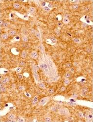
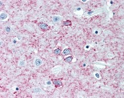
- Immunohistochemistry-Paraffin: Adenosine A2aR Antibody (7F6-G5-A2) [NBP1-39474] - Analysis of a FFPE tissue section of mouse brain using Adenosine A2a R antibody (clone 7F6-G5-A2) at 1:400 dilution. The antibody generated nice membranous / punctate staining of Adenosine A2a receptors.

- Submitted by
- Novus Biologicals (provider)
- Main image
- Experimental details
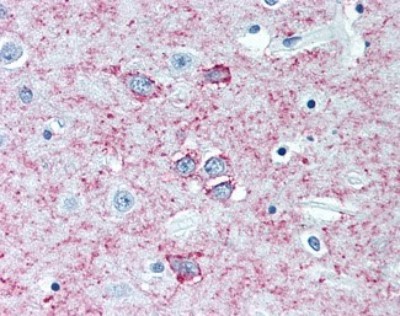
- Immunohistochemistry-Paraffin: Adenosine A2aR Antibody (7F6-G5-A2) [NBP1-39474] - Analysis of a FFPE tissue section of mouse brain using Adenosine A2a R antibody (clone 7F6-G5-A2) at 1:100 dilution. The antibody generated nice membranous / punctate staining of Adenosine A2a receptors.

- Submitted by
- Novus Biologicals (provider)
- Main image
- Experimental details
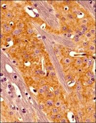
- Immunohistochemistry-Paraffin: Adenosine A2aR Antibody (7F6-G5-A2) [NBP1-39474] - Staining of human brain (putamen), antibody at 5 ug/mL.
Supportive validation
- Submitted by
- Novus Biologicals (provider)
- Main image
- Experimental details
- Flow (Intracellular): Adenosine A2aR Antibody (7F6-G5-A2) [NBP1-39474] - An intracellular stain was performed on hPBMCs with Adenosine A2a R (7F6-G5-A2) antibody NBP1-39474 and a matched isotype control NBP2-14864. Cells were fixed with 4% PFA and then permeablized with 0.1% saponin. Cells were incubated in an antibody dilution of 1 ug/mL for 30 minutes at room temperature, followed by mouse F(ab)2 IgG (H+L) APC-conjugated secondary antibody (F0101B, R&D Systems).
- Submitted by
- Novus Biologicals (provider)
- Main image
- Experimental details
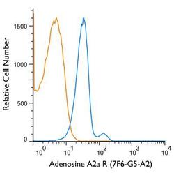
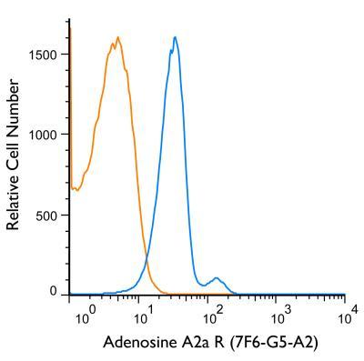
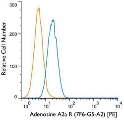
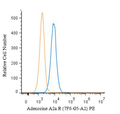
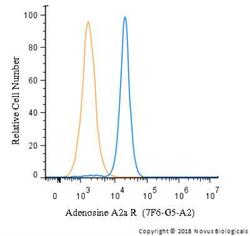
- Flow Cytometry: Adenosine A2aR Antibody (7F6-G5-A2) [NBP1-39474] - Using the PE direct conjugate An intracellular stain was performed on SH-SY5Y cells with Adenosine A2a R (7F6-G5-A2) antibody NBP1-39474PE (blue) and a matched isotype control NB600-986PE (orange). Cells were fixed with 4% PFA and then permeablized with 0.1% saponin. Cells were incubated in an antibody dilution of 1 ug/mL for 30 minutes at room temperature. Both antibodies were conjugated to Phycoerythrin (PE).

- Submitted by
- Novus Biologicals (provider)
- Main image
- Experimental details
- Flow (Intracellular): Adenosine A2aR Antibody (7F6-G5-A2) [NBP1-39474] - An intracellular stain was performed on U-937 cells with Adenosine A2a R (7F6-G5-A2) antibody NBP1-39474 and a matched isotype control. Cells were fixed with 4% PFA and then permeablized with 0.1% saponin. Cells were incubated in an antibody dilution of 2.5 ug/mL for 30 minutes at room temperature, followed by mouse F(ab)2 IgG (H+L) PE-conjugated secondary antibody (F0102B, R&D Systems).
- Submitted by
- Novus Biologicals (provider)
- Main image
- Experimental details
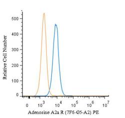
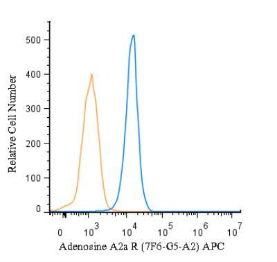
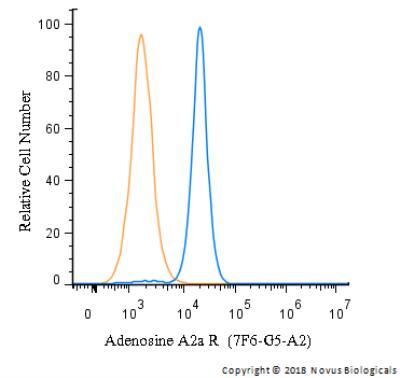
- Flow (Intracellular): Adenosine A2aR Antibody (7F6-G5-A2) [NBP1-39474] - An intracellular stain was performed on U-937 cells with Adenosine A2a R (7F6-G5-A2) antibody NBP1-39474PE (blue) and a matched isotype control (orange). Cells were fixed with 4% PFA and then permeablized with 0.1% saponin. Cells were incubated in an antibody dilution of 5 ug/mL for 30 minutes at room temperature. Both antibodies were conjugated to Phycoerythrin (PE).
- Submitted by
- Novus Biologicals (provider)
- Main image
- Experimental details
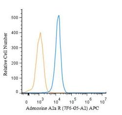
- Flow (Intracellular): Adenosine A2aR Antibody (7F6-G5-A2) [NBP1-39474] - An intracellular stain was performed on U-937 cells with Adenosine A2a R (7F6-G5-A2) antibody NBP1-39474APC (blue) and a matched isotype control (orange). Cells were fixed with 4% PFA and then permeablized with 0.1% saponin. Cells were incubated in an antibody dilution of 1 ug/mL for 30 minutes at room temperature. Both antibodies were conjugated to Allophycocyanin (APC).
- Submitted by
- Novus Biologicals (provider)
- Main image
- Experimental details
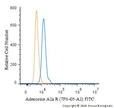
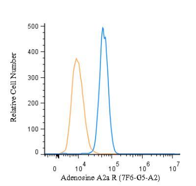
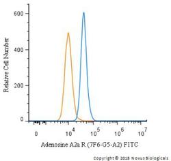
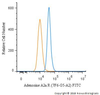
- Flow Cytometry: Adenosine A2aR Antibody (7F6-G5-A2) [NBP1-39474] - An intracellular stain was performed on U-87 MG cells with Adenosine A2a R (7F6-G5-A2) antibody NBP1-39474F (blue) and a matched isotype control (orange). Cells were fixed with 4% PFA and then permeablized with 0.1% saponin. Cells were incubated in an antibody dilution of 10 ug/mL for 30 minutes at room temperature. Both antibodies were conjugated to FITC.
- Submitted by
- Novus Biologicals (provider)
- Main image
- Experimental details
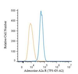
- Flow Cytometry: Adenosine A2aR Antibody (7F6-G5-A2) [NBP1-39474] - An intracellular stain was performed on SH-SY5Y with Adenosine A2a R Antibody (7F6-G5-A2) NBP1-39474 and a matched isotype control. Cells were fixed with 4% PFA and then permeablized with 0.1% saponin. Cells were incubated in an antibody dilution of 1 ug/mL for 30 minutes at room temperature, followed by Mouse F(ab)2 IgG (H+L) PE-conjugated Antibody (R&D Systems, F0102B).
- Submitted by
- Novus Biologicals (provider)
- Main image
- Experimental details
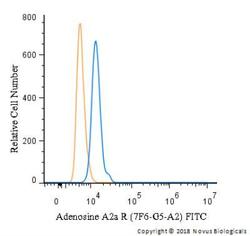
- Flow Cytometry: Adenosine A2aR Antibody (7F6-G5-A2) [NBP1-39474] - An intracellular stain was performed on U-937 cells with Adenosine A2a R (7F6-G5-A2) antibody NBP1-39474F (blue) and a matched isotype control (orange). Cells were fixed with 4% PFA and then permeablized with 0.1% saponin. Cells were incubated in an antibody dilution of 5 ug/mL for 30 minutes at room temperature. Both antibodies were conjugated to FITC.