Explore
 Validate
 Learn
Learn Western blot
 ELISA
ELISA Immunocytochemistry
Antibody data
- Antibody Data
- Antigen structure
- References [19]
- Comments [0]
- Validations
- Immunocytochemistry [1]
- Immunohistochemistry [2]
- Other assay [10]
Submit
Validation data
Reference
Comment
Report error
- Product number
- 13-4400 - Provider product page

- Provider
- Invitrogen Antibodies
- Product name
- Perlecan Monoclonal Antibody (7B5)
- Antibody type
- Monoclonal
- Antigen
- Other
- Reactivity
- Human
- Host
- Mouse
- Isotype
- IgG
- Antibody clone number
- 7B5
- Vial size
- 100 μg
- Concentration
- 0.5 mg/mL
- Storage
- -20°C
Submitted references Evaluation of Human-Induced Pluripotent Stem Cells Derived from a Patient with Schwartz-Jampel Syndrome Revealed Distinct Hyperexcitability in the Skeletal Muscles.
Human blood vessel organoids as a model of diabetic vasculopathy.
Influence of fibrin matrices and their released factors on epidermal substitute phenotype and engraftment.
Bronchial extracellular matrix from COPD patients induces altered gene expression in repopulated primary human bronchial epithelial cells.
Effects of a skin-massaging device on the ex-vivo expression of human dermis proteins and in-vivo facial wrinkles.
Remodeling of extracellular matrix by normal and tumor-associated fibroblasts promotes cervical cancer progression.
Peripheral nerve hyperexcitability with preterminal nerve and neuromuscular junction remodeling is a hallmark of Schwartz-Jampel syndrome.
Confluence switch signaling regulates ECM composition and the plasmin proteolytic cascade in keratinocytes.
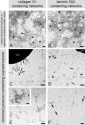
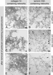
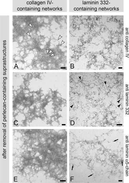
The epidermal basement membrane is a composite of separate laminin- or collagen IV-containing networks connected by aggregated perlecan, but not by nidogens.
Similarity of recombinant human perlecan domain 1 by alternative expression systems bioactive heterogenous recombinant human perlecan D1.
Quantitative and qualitative alterations of heparan sulfate in fibrogenic liver diseases and hepatocellular cancer.
Peroxynitrite modifies the structure and function of the extracellular matrix proteoglycan perlecan by reaction with both the protein core and the heparan sulfate chains.
Vascular gene expression patterns are conserved in primary and metastatic brain tumors.
Pericyte recruitment during vasculogenic tube assembly stimulates endothelial basement membrane matrix formation.
Differences in heparan sulfate production in cervical fibroblast cultures from women undergoing term and preterm delivery.
The structure, location, and function of perlecan, a prominent pericellular proteoglycan of fetal, postnatal, and mature hyaline cartilages.
Primary human glomerular endothelial cells produce proteoglycans, and puromycin affects their posttranslational modification.
Atypical development of the tracheal basement membrane zone of infant rhesus monkeys exposed to ozone and allergen.
Fibroblast growth factor-2 during postnatal development of the tracheal basement membrane zone.
Yamashita Y, Nakada S, Nakamura K, Sakurai H, Ohno K, Goto T, Mabuchi Y, Akazawa C, Hattori N, Arikawa-Hirasawa E
Biomedicines 2023 Mar 7;11(3)
Biomedicines 2023 Mar 7;11(3)
Human blood vessel organoids as a model of diabetic vasculopathy.
Wimmer RA, Leopoldi A, Aichinger M, Wick N, Hantusch B, Novatchkova M, Taubenschmid J, Hämmerle M, Esk C, Bagley JA, Lindenhofer D, Chen G, Boehm M, Agu CA, Yang F, Fu B, Zuber J, Knoblich JA, Kerjaschki D, Penninger JM
Nature 2019 Jan;565(7740):505-510
Nature 2019 Jan;565(7740):505-510
Influence of fibrin matrices and their released factors on epidermal substitute phenotype and engraftment.
Alexaline MM, Magne B, Zuleta Rodríguez A, Nivet M, Bacqueville D, Lataillade JJ, Trouillas M
Journal of tissue engineering and regenerative medicine 2019 Aug;13(8):1362-1374
Journal of tissue engineering and regenerative medicine 2019 Aug;13(8):1362-1374
Bronchial extracellular matrix from COPD patients induces altered gene expression in repopulated primary human bronchial epithelial cells.
Hedström U, Hallgren O, Öberg L, DeMicco A, Vaarala O, Westergren-Thorsson G, Zhou X
Scientific reports 2018 Feb 22;8(1):3502
Scientific reports 2018 Feb 22;8(1):3502
Effects of a skin-massaging device on the ex-vivo expression of human dermis proteins and in-vivo facial wrinkles.
Caberlotto E, Ruiz L, Miller Z, Poletti M, Tadlock L
PloS one 2017;12(3):e0172624
PloS one 2017;12(3):e0172624
Remodeling of extracellular matrix by normal and tumor-associated fibroblasts promotes cervical cancer progression.
Fullár A, Dudás J, Oláh L, Hollósi P, Papp Z, Sobel G, Karászi K, Paku S, Baghy K, Kovalszky I
BMC cancer 2015 Apr 11;15:256
BMC cancer 2015 Apr 11;15:256
Peripheral nerve hyperexcitability with preterminal nerve and neuromuscular junction remodeling is a hallmark of Schwartz-Jampel syndrome.
Bauché S, Boerio D, Davoine CS, Bernard V, Stum M, Bureau C, Fardeau M, Romero NB, Fontaine B, Koenig J, Hantaï D, Gueguen A, Fournier E, Eymard B, Nicole S
Neuromuscular disorders : NMD 2013 Dec;23(12):998-1009
Neuromuscular disorders : NMD 2013 Dec;23(12):998-1009
Confluence switch signaling regulates ECM composition and the plasmin proteolytic cascade in keratinocytes.
Botta A, Delteil F, Mettouchi A, Vieira A, Estrach S, Négroni L, Stefani C, Lemichez E, Meneguzzi G, Gagnoux-Palacios L
Journal of cell science 2012 Sep 15;125(Pt 18):4241-52
Journal of cell science 2012 Sep 15;125(Pt 18):4241-52
The epidermal basement membrane is a composite of separate laminin- or collagen IV-containing networks connected by aggregated perlecan, but not by nidogens.
Behrens DT, Villone D, Koch M, Brunner G, Sorokin L, Robenek H, Bruckner-Tuderman L, Bruckner P, Hansen U
The Journal of biological chemistry 2012 May 25;287(22):18700-9
The Journal of biological chemistry 2012 May 25;287(22):18700-9
Similarity of recombinant human perlecan domain 1 by alternative expression systems bioactive heterogenous recombinant human perlecan D1.
Ellis AL, Pan W, Yang G, Jones K, Chuang C, Whitelock JM, DeCarlo AA
BMC biotechnology 2010 Sep 9;10:66
BMC biotechnology 2010 Sep 9;10:66
Quantitative and qualitative alterations of heparan sulfate in fibrogenic liver diseases and hepatocellular cancer.
Tátrai P, Egedi K, Somorácz A, van Kuppevelt TH, Ten Dam G, Lyon M, Deakin JA, Kiss A, Schaff Z, Kovalszky I
The journal of histochemistry and cytochemistry : official journal of the Histochemistry Society 2010 May;58(5):429-41
The journal of histochemistry and cytochemistry : official journal of the Histochemistry Society 2010 May;58(5):429-41
Peroxynitrite modifies the structure and function of the extracellular matrix proteoglycan perlecan by reaction with both the protein core and the heparan sulfate chains.
Kennett EC, Rees MD, Malle E, Hammer A, Whitelock JM, Davies MJ
Free radical biology & medicine 2010 Jul 15;49(2):282-93
Free radical biology & medicine 2010 Jul 15;49(2):282-93
Vascular gene expression patterns are conserved in primary and metastatic brain tumors.
Liu Y, Carson-Walter EB, Cooper A, Winans BN, Johnson MD, Walter KA
Journal of neuro-oncology 2010 Aug;99(1):13-24
Journal of neuro-oncology 2010 Aug;99(1):13-24
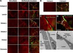
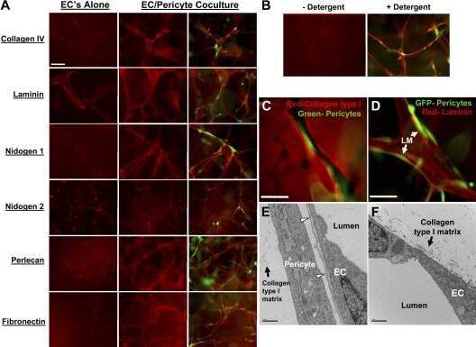
Pericyte recruitment during vasculogenic tube assembly stimulates endothelial basement membrane matrix formation.
Stratman AN, Malotte KM, Mahan RD, Davis MJ, Davis GE
Blood 2009 Dec 3;114(24):5091-101
Blood 2009 Dec 3;114(24):5091-101
Differences in heparan sulfate production in cervical fibroblast cultures from women undergoing term and preterm delivery.
Akerud A, Dubicke A, Sennstrom M, Ekman-Ordeberg G, Malmstrom A
Acta obstetricia et gynecologica Scandinavica 2008;87(11):1220-8
Acta obstetricia et gynecologica Scandinavica 2008;87(11):1220-8
The structure, location, and function of perlecan, a prominent pericellular proteoglycan of fetal, postnatal, and mature hyaline cartilages.
Melrose J, Roughley P, Knox S, Smith S, Lord M, Whitelock J
The Journal of biological chemistry 2006 Dec 1;281(48):36905-14
The Journal of biological chemistry 2006 Dec 1;281(48):36905-14
Primary human glomerular endothelial cells produce proteoglycans, and puromycin affects their posttranslational modification.
Björnson A, Moses J, Ingemansson A, Haraldsson B, Sörensson J
American journal of physiology. Renal physiology 2005 Apr;288(4):F748-56
American journal of physiology. Renal physiology 2005 Apr;288(4):F748-56
Atypical development of the tracheal basement membrane zone of infant rhesus monkeys exposed to ozone and allergen.
Evans MJ, Fanucchi MV, Baker GL, Van Winkle LS, Pantle LM, Nishio SJ, Schelegle ES, Gershwin LJ, Miller LA, Hyde DM, Sannes PL, Plopper CG
American journal of physiology. Lung cellular and molecular physiology 2003 Oct;285(4):L931-9
American journal of physiology. Lung cellular and molecular physiology 2003 Oct;285(4):L931-9
Fibroblast growth factor-2 during postnatal development of the tracheal basement membrane zone.
Evans MJ, Fanucchi MV, Van Winkle LS, Baker GL, Murphy AE, Nishio SJ, Sannes PL, Plopper CG
American journal of physiology. Lung cellular and molecular physiology 2002 Dec;283(6):L1263-70
American journal of physiology. Lung cellular and molecular physiology 2002 Dec;283(6):L1263-70
No comments: Submit comment
Supportive validation
- Submitted by
- Invitrogen Antibodies (provider)
- Main image
- Experimental details
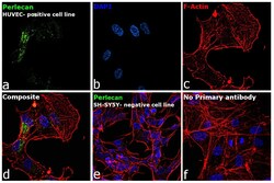
- Immunofluorescence analysis of HSPG2 was performed using 70% confluent log phase HUVEC and SH-SY5Y cells. The cells were fixed with 4% paraformaldehyde for 10 minutes, permeabilized with 0.1% Triton™ X-100 for 15 minutes, and blocked with 2% BSA for 45 minutes at room temperature. The cells were labeled with Perlecan Monoclonal Antibody (7B5) (Product # 13-4400, 1:100) in 0.1% BSA, incubated at 4 degree celsius overnight and then labeled with Donkey anti-Mouse IgG (H+L) Highly Cross-Adsorbed Secondary Antibody, Alexa Fluor™ Plus 488 (Product # A32766, 1:2,000), for 45 minutes at room temperature (Panel a: Green). Nuclei (Panel b: Blue) were stained with ProLong™ Diamond Antifade Mountant with DAPI (Product # P36962). F-actin (Panel c: Red) was stained with Rhodamine Phalloidin (Product # R415, 1:300). Panel d represents the merged image showing golgi-like localization. Panel e represents SH-SY5Y cells with lower HSPG2 expression. Panel f represents control cells with no primary antibody to assess background. The images were captured at 60X magnification.

Supportive validation
- Submitted by
- Invitrogen Antibodies (provider)
- Main image
- Experimental details
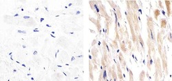
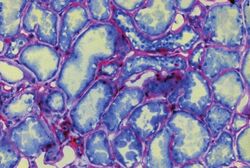
- Immunohistochemistry analysis of PERLECAN showing staining in the cytoplasm of paraffin-embedded human heart tissue (right) compared to a negative control without primary antibody (left). To expose target proteins, antigen retrieval was performed using 10mM sodium citrate (pH 6.0), microwaved for 8-15 min. Following antigen retrieval, tissues were blocked in 3% H2O2-methanol for 15 min at room temperature, washed with ddH2O and PBS, and then probed with a PERLECAN Mouse Monoclonal Antibody (Product # 13-4400) diluted in 3% BSA-PBS at a dilution of 1:20 overnight at 4°C in a humidified chamber. Tissues were washed extensively in PBST and detection was performed using an HRP-conjugated secondary antibody followed by colorimetric detection using a DAB kit. Tissues were counterstained with hematoxylin and dehydrated with ethanol and xylene to prep for mounting.
- Submitted by
- Invitrogen Antibodies (provider)
- Main image
- Experimental details
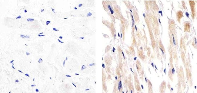
- Immunohistochemical staining using mouse anti-perlecan monoclonal antibody (Product # 13-4400).

Supportive validation
- Submitted by
- Invitrogen Antibodies (provider)
- Main image
- Experimental details
- NULL
- Submitted by
- Invitrogen Antibodies (provider)
- Main image
- Experimental details
- NULL

- Submitted by
- Invitrogen Antibodies (provider)
- Main image
- Experimental details
- NULL
- Submitted by
- Invitrogen Antibodies (provider)
- Main image

- Experimental details
- NULL
- Submitted by
- Invitrogen Antibodies (provider)
- Main image
- Experimental details
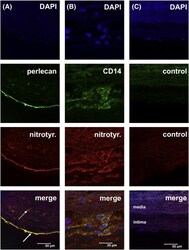
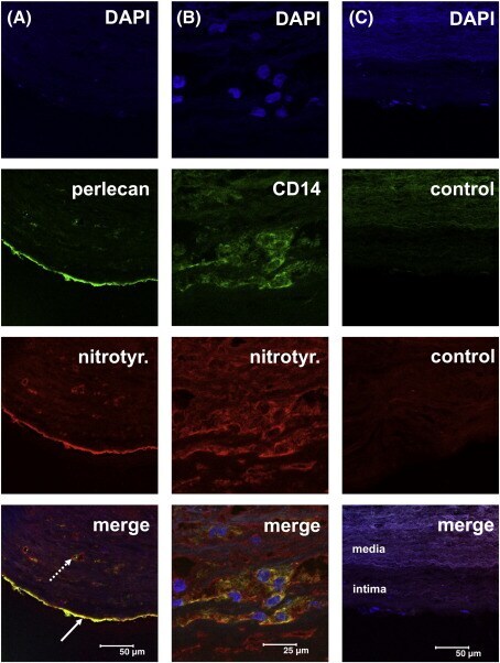
- Fig. 10 Double immunofluorescence staining for perlecan, 3-nitrotyrosine epitopes and CD14-positive cells in human atherosclerotic lesion sections. Frozen sections (5 mum) of human atherosclerotic lesions were incubated with monoclonal or polyclonal primary antibodies: anti-nitrotyrosine (rabbit IgG), anti-human perlecan (mouse mAb, clone 7B5, perlecan domain III) anti-human CD14 (mouse mAb), non-immune rabbit IgG and non-immune mouse IgG, and subsequently the following detection antibodies: goat anti-rabbit cyanine-3 (Cy-3)-labelled IgG or goat anti-mouse Cy-2-labeled IgG. DAPI was used to image cell nuclei. Images were acquired as described in the Materials and Methods. A) Heavily thickened intima of an artery with a type III/IV lesion. Epitopes for 3-nitrotyrosine (red signal) show marked co-localization with perlecan (green) present in the basement membrane of the endothelium (arrow) as well as with those of the vasa vasorum (dotted arrow). B) The signal for 3-nitrotyrosine (red) was frequently co-localized with CD14-positive cells (macrophages, green). The red signal for nitrotyrosine underneath the bulk of macrophages results from multiple oblique sections through a vas vasorum. C) In sections where the antibodies for perlecan/CD14 and for 3-nitrotyrosine were replaced with non-immune mouse IgG (control, green) and non-immune rabbit IgG (control, red), no staining in the intima was observed. The faint background staining in the media (also observed when no primary or se
- Submitted by
- Invitrogen Antibodies (provider)
- Main image
- Experimental details
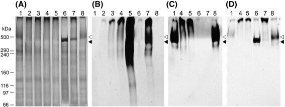
- Fig. 5 Structural consequences of perlecan modification by peroxynitrite. Perlecan (330 nM) in 0.1 M phosphate buffer, pH 7 (lane 1) was exposed for 20 min at 22 degC to peroxynitrite at molar ratios of 250 (lane 2), 500 (lane 3), 1000 (lane 4), 1000 in the presence of 25 mM bicarbonate (lane 5), heparinase III for 16 h at 37 degC (lane 6), heparinase III followed by 1000-fold molar excess of peroxynitrite (lane 7), and dONOO (lane 8), prior to separation on 3-8% Tris-acetate gels under reducing conditions for 1 h at 150 V and subsequent western blotting to nitrocellulose. (A) Stains-all/silver stain of gel. (B) Western blot probed for 3-nitroTyr formation with mAb against 3-nitroTyr (HM11). (C) Western blot probed for heparan sulfate epitopes with mAb 10E4. (D) Western blot probed for perlecan domain III with mAb 7B5. Position of molecular mass markers are shown for reference: < perlecan heparan sulfate, < perlecan protein core.
- Submitted by
- Invitrogen Antibodies (provider)
- Main image
- Experimental details
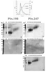
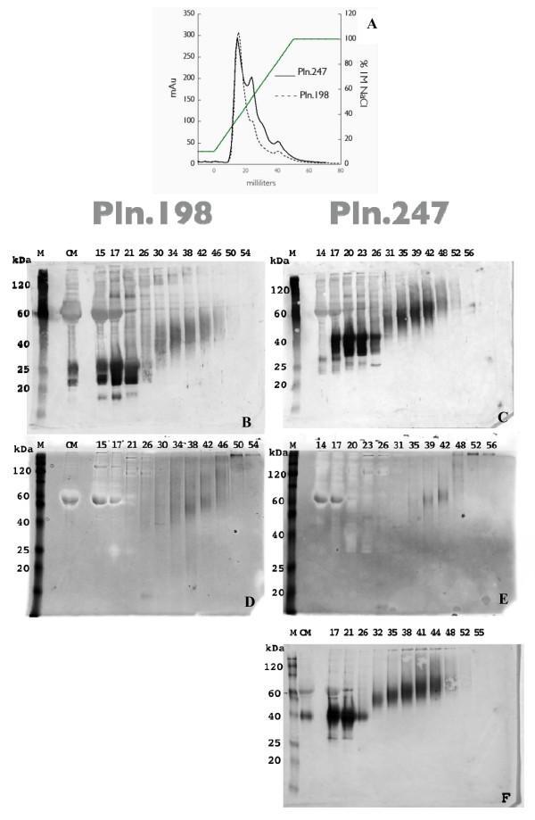
- Figure 1 Anion Exchange Chromatography of rhPln.247 conditioned media (CM) . A: Chromatogram of elution profile for rhPln.198-Ad and rhPln.247-Ad CM expressed from HEK 293 cells; B and C: Western blots of anion exchange fractions with mAb CSI 001-71 recognizing the Pln.D1 core; D and E: Stains-All analysis of HEK 293 fractions; F: Fractions from HUVEC synthesis. Panels B and D represent rhPln.198 fractionation; panels C, E, F represent rhPln.247 fractionation. (M) Markers; (CM) Conditioned medium undiluted from cells. Similar data were generated using plasmid expression of pln.247 (not shown).
- Submitted by
- Invitrogen Antibodies (provider)
- Main image
- Experimental details
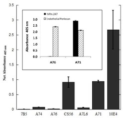
- Figure 2 ELISA identifies only Pln.D1 but not Pln.D3-5 in enriched rhPln.198 . The enriched high M r rhPln.198 synthesized by HEK 293 cells was used to coat microtiter wells that were subsequently blocked then incubated with 1.7 mug/ml of different primary antibodies as depicted in the legend. ELISA was completed as described in Methods. Background signal for each antibody binding to uncoated wells was subtracted to produce the net signal. Inset: ELISA data showing negligible mAb A76 reactivity against rhPln.247 and strong reactivity with full-length endothelial perlecan (purified as previously described [ 53 ]), while mAb A71 reacted strongly to both the native perlecan and the truncated recombinant. Net absorbance minus buffer-coated wells represented.
- Submitted by
- Invitrogen Antibodies (provider)
- Main image

- Experimental details
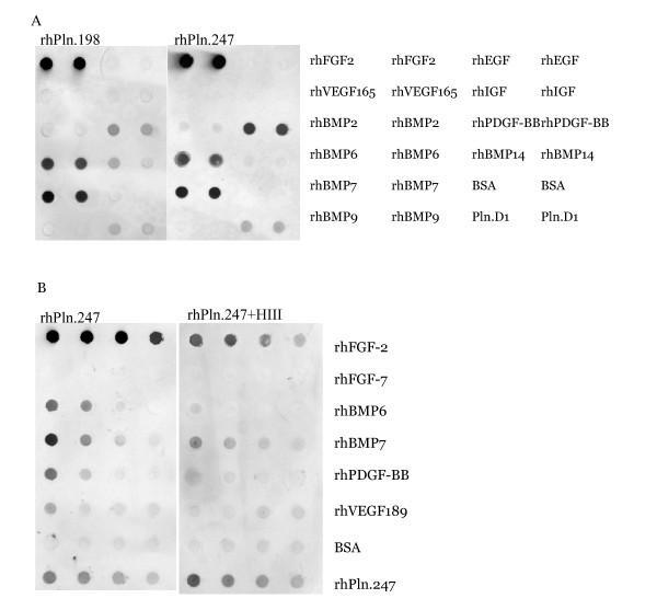
- Figure 8 Immunoblot analysis of growth factor binding to Pln.198 and Pln.247 . 500 ng growth factors were bound to nitrocellulose, the blots were blocked, and then incubated in either 1 ug/ml Pln.198 or Pln.247 (panel A) or rhPln.247 pre-digested with heparinase III (panel B). Detection was performed with anti-perlecan domain 1 primary antibody CSI 001-71. Note that while both the background BSA signal and the internal control rhPln.247 signals are slightly higher in panel B (bottom two samples), a decrease in the growth factor binding to Pln.247 was present after digestion.
- Submitted by
- Invitrogen Antibodies (provider)
- Main image
- Experimental details
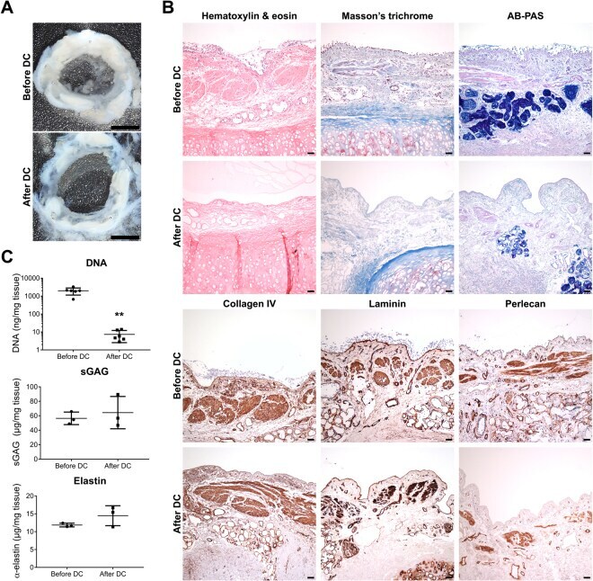
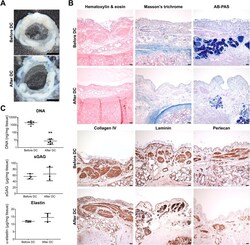
- Figure 2 Decellularization of human bronchial airways efficiently removes cells while preserving the extracellular matrix. ( A) The macroscopic structure of the 500 um thick human bronchial tissue sections was preserved after decellularization (DC). (B) Hematoxylin & eosin, Masson's trichrome (collagen in blue) and alcian-blue periodic acid Schiff (AB-PAS) (polysaccharides in magenta/purple) stainings as well as immunohistochemistry against the basement membrane proteins collagen IV, laminin and perlecan, before and after DC. Images are representative of n = 3. (C) Dye-binding methods confirmed that DC efficiently decreased DNA content (**p = 0.002) while preserving sulfated glycosaminoglycans (sGAG) and elastin in bronchial scaffolds (n = 6 for DNA, n = 3 for sGAG and elastin). The data were analyzed with the Mann-Whitney test and graphs indicate mean and standard deviation. Scale bars: 3 mm in A and 50 um in B.