Explore
 Validate
 Learn
Learn Western blot
Antibody data
- Antibody Data
- Antigen structure
- References [10]
- Comments [0]
- Validations
- Western blot [1]
- Immunohistochemistry [1]
- Flow cytometry [3]
Submit
Validation data
Reference
Comment
Report error
- Product number
- MA5-14413 - Provider product page

- Provider
- Invitrogen Antibodies
- Product name
- CD38 Monoclonal Antibody (38C03 (SPC32))
- Antibody type
- Monoclonal
- Antigen
- Recombinant full-length protein
- Description
- MA5-14413 targets CD38 in WB, FACS and IHC (P) applications and shows reactivity with Human and Mouse samples. This antibody is not suitable for Jurkat cell lysates or mouse thymus tissues in Western blot analysis.
- Antibody clone number
- 38C03 (SPC32)
- Concentration
- Conc. Not Determined
Submitted references Unusual bilateral kidney and duodenal plasmablastic lymphoma presentation in an elderly patient: A case report.
TBL1XR1 Mutations Drive Extranodal Lymphoma by Inducing a Pro-tumorigenic Memory Fate.
Plasma cells within granulomatous inflammation display signs pointing to autoreactivity and destruction in granulomatosis with polyangiitis.
VR09 cell line: an EBV-positive lymphoblastoid cell line with in vivo characteristics of diffuse large B cell lymphoma of activated B-cell type.
Upregulation of tumor necrosis factor receptor-associated factor 6 correlated with synovitis severity in rheumatoid arthritis.
Reconstructing the human hematopoietic niche in immunodeficient mice: opportunities for studying primary multiple myeloma.
Analysis of the IgVH genes in T cell-mediated and antibody-mediated rejection of the kidney graft.
Diagnostic and differential diagnostic criteria of lymphoid neoplasms in bone marrow trephine biopsies: a study of 87 cases.
Channelling of substrate promiscuity of the skeletal-muscle ADP-ribosyl cyclase isoform.
Human adult craniofacial muscle-derived cells: neural-cell adhesion-molecule (NCAM; CD56)-expressing cells appear to contain multipotential stem cells.
Liu YC, Su YT, Huang CK, Tsai YC, Chen YC, Li PF
Molecular and clinical oncology 2022 Jul;17(1):122
Molecular and clinical oncology 2022 Jul;17(1):122
TBL1XR1 Mutations Drive Extranodal Lymphoma by Inducing a Pro-tumorigenic Memory Fate.
Venturutti L, Teater M, Zhai A, Chadburn A, Babiker L, Kim D, Béguelin W, Lee TC, Kim Y, Chin CR, Yewdell WT, Raught B, Phillip JM, Jiang Y, Staudt LM, Green MR, Chaudhuri J, Elemento O, Farinha P, Weng AP, Nissen MD, Steidl C, Morin RD, Scott DW, Privé GG, Melnick AM
Cell 2020 Jul 23;182(2):297-316.e27
Cell 2020 Jul 23;182(2):297-316.e27
Plasma cells within granulomatous inflammation display signs pointing to autoreactivity and destruction in granulomatosis with polyangiitis.
Mueller A, Brieske C, Schinke S, Csernok E, Gross WL, Hasselbacher K, Voswinkel J, Holl-Ulrich K
Arthritis research & therapy 2014 Feb 20;16(1):R55
Arthritis research & therapy 2014 Feb 20;16(1):R55
VR09 cell line: an EBV-positive lymphoblastoid cell line with in vivo characteristics of diffuse large B cell lymphoma of activated B-cell type.
Nichele I, Zamò A, Bertolaso A, Bifari F, Tinelli M, Franchini M, Stradoni R, Aprili F, Pizzolo G, Krampera M
PloS one 2012;7(12):e52811
PloS one 2012;7(12):e52811
Upregulation of tumor necrosis factor receptor-associated factor 6 correlated with synovitis severity in rheumatoid arthritis.
Zhu LJ, Dai L, Zheng DH, Mo YQ, Ou-Yang X, Wei XN, Shen J, Zhang BY
Arthritis research & therapy 2012 Jun 4;14(3):R133
Arthritis research & therapy 2012 Jun 4;14(3):R133
Reconstructing the human hematopoietic niche in immunodeficient mice: opportunities for studying primary multiple myeloma.
Groen RW, Noort WA, Raymakers RA, Prins HJ, Aalders L, Hofhuis FM, Moerer P, van Velzen JF, Bloem AC, van Kessel B, Rozemuller H, van Binsbergen E, Buijs A, Yuan H, de Bruijn JD, de Weers M, Parren PW, Schuringa JJ, Lokhorst HM, Mutis T, Martens AC
Blood 2012 Jul 19;120(3):e9-e16
Blood 2012 Jul 19;120(3):e9-e16
Analysis of the IgVH genes in T cell-mediated and antibody-mediated rejection of the kidney graft.
Bellan C, Amato T, Carmellini M, Onorati M, D'Amuri A, Leoncini L, del Vecchio MT
Journal of clinical pathology 2011 Jan;64(1):47-53
Journal of clinical pathology 2011 Jan;64(1):47-53
Diagnostic and differential diagnostic criteria of lymphoid neoplasms in bone marrow trephine biopsies: a study of 87 cases.
Horváth E, Mezei T, Pávai Z, Turcu M, Demian S, Tóth E, Chira L, Jung I
Romanian journal of morphology and embryology = Revue roumaine de morphologie et embryologie 2009;50(3):399-406
Romanian journal of morphology and embryology = Revue roumaine de morphologie et embryologie 2009;50(3):399-406
Channelling of substrate promiscuity of the skeletal-muscle ADP-ribosyl cyclase isoform.
Bacher I, Zidar A, Kratzel M, Hohenegger M
The Biochemical journal 2004 Jul 1;381(Pt 1):147-54
The Biochemical journal 2004 Jul 1;381(Pt 1):147-54
Human adult craniofacial muscle-derived cells: neural-cell adhesion-molecule (NCAM; CD56)-expressing cells appear to contain multipotential stem cells.
Sinanan AC, Hunt NP, Lewis MP
Biotechnology and applied biochemistry 2004 Aug;40(Pt 1):25-34
Biotechnology and applied biochemistry 2004 Aug;40(Pt 1):25-34
No comments: Submit comment
Supportive validation
- Submitted by
- Invitrogen Antibodies (provider)
- Main image
- Experimental details
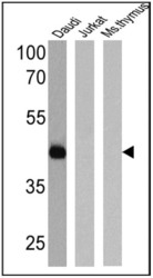
- Western blot analysis of CD38 was performed by loading 25 µg of Daudi (Lane 1), Jurkat (Lane 2), and mouse thymus cell lysates (Lane 3) and a molecular weight protein ladder onto an SDS polyacrylamide gel. Proteins were transferred to a PVDF membrane and blocked with a blocking buffer at 4ºC overnight. The membrane was probed with a CD38 monoclonal antibody (Product # MA5-14413) at a dilution of 1:50 overnight at 4°C, washed in TBST, and probed with an HRP-conjugated secondary antibody for 1 hr at room temperature in the dark. Chemiluminescent detection was performed using Pierce ECL Plus Western Blotting Substrate (Product # 32132). Results show a band at 45 kDa in Daudi cells only.

Supportive validation
- Submitted by
- Invitrogen Antibodies (provider)
- Main image
- Experimental details
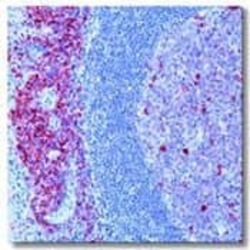
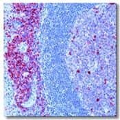
- Formalin-fixed, paraffin-embedded human tonsil stained with CD38 using peroxidase-conjugate and AEC chromogen. Note membrane staining of lymphocytes.
Supportive validation
- Submitted by
- Invitrogen Antibodies (provider)
- Main image

- Experimental details
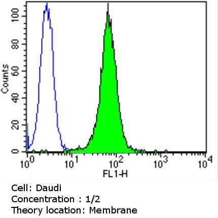
- Flow cytometry analysis of CD38 in Daudi cells (green) compared to an isotype control (blue). Cells were harvested, adjusted to a concentration of 1-5x10^6 cells/mL, fixed with 2% paraformaldehyde and washed with PBS. Cells were blocked with a 2% solution of BSA-PBS for 30 min at room temperature and incubated with a CD38 monoclonal antibody (Product # MA5-14413) at a dilution of 1:2 for 60 min at room temperature. Cells were then incubated for 40 min at room temperature in the dark using a Dylight 488-conjugated goat anti-mouse IgG (H+L) secondary antibody and re-suspended in PBS for FACS analysis.
- Submitted by
- Invitrogen Antibodies (provider)
- Main image
- Experimental details
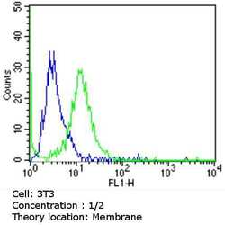
- Flow cytometry analysis of CD38 in NIH-3T3 cells (green) compared to an isotype control (blue). Cells were harvested, adjusted to a concentration of 1-5x10^6 cells/mL, fixed with 2% paraformaldehyde and washed with PBS. Cells were blocked with a 2% solution of BSA-PBS for 30 min at room temperature and incubated with a CD38 monoclonal antibody (Product # MA5-14413) at a dilution of 1:2 for 60 min at room temperature. Cells were then incubated for 40 min at room temperature in the dark using a Dylight 488-conjugated goat anti-mouse IgG (H+L) secondary antibody and re-suspended in PBS for FACS analysis.

- Submitted by
- Invitrogen Antibodies (provider)
- Main image
- Experimental details
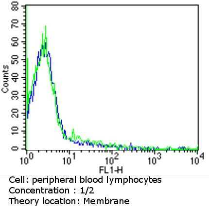
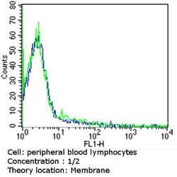
- Flow cytometry analysis of CD38 in PBMC cells (green) compared to an isotype control (blue). Human blood was collected, combined with a hydrophilic polysaccharide, centrifuged, transferred to a conical tube and washed with PBS. 50 µL of cell solution was added to each tube at a dilution of 2x10^7 cells/mL, followed by the addition of 50 µL of isotype control and primary antibody (Product # MA5-14413) at a dilution of 1:2. Cells were incubated for 30 min at 4ºC and washed with a cell buffer, followed by incubation with a DyLight 488-conjugated goat anti-mouse IgG (H+L) secondary for 30 min at 4ºC in the dark. FACS analysis was performed using 400 µL of cell buffer.