Explore
 Validate
 Learn
Learn Western blot
 Immunohistochemistry
Antibody data
- Antibody Data
- Antigen structure
- References [3]
- Comments [0]
- Validations
- Immunohistochemistry [1]
- Other assay [4]
Submit
Validation data
Reference
Comment
Report error
- Product number
- PA5-27165 - Provider product page

- Provider
- Invitrogen Antibodies
- Product name
- ADAMTS5 Polyclonal Antibody
- Antibody type
- Polyclonal
- Antigen
- Synthetic peptide
- Description
- Recommended positive controls: HCT116, MCF-7, JC. Predicted reactivity: Mouse (100%), Rat (100%), Bovine (100%). Store product as a concentrated solution. Centrifuge briefly prior to opening the vial.
- Reactivity
- Human, Mouse
- Host
- Rabbit
- Isotype
- IgG
- Vial size
- 100 μL
- Concentration
- 1 mg/mL
- Storage
- Store at 4°C short term. For long term storage, store at -20°C, avoiding freeze/thaw cycles.
Submitted references Bone Marrow Mesenchymal Stem Cell-Derived Extracellular Vesicles Carrying circ_0050205 Attenuate Intervertebral Disc Degeneration.
Downregulation of MicroRNA-495 Alleviates IL-1β Responses among Chondrocytes by Preventing SOX9 Reduction.
Galnt1 is required for normal heart valve development and cardiac function.
Yu XJ, Liu QK, Lu R, Wang SX, Xu HR, Wang YG, Bao Y, Jiang YQ, Li MW, Kang H
Oxidative medicine and cellular longevity 2022;2022:8983667
Oxidative medicine and cellular longevity 2022;2022:8983667
Downregulation of MicroRNA-495 Alleviates IL-1β Responses among Chondrocytes by Preventing SOX9 Reduction.
Joung S, Yoon DS, Cho S, Ko EA, Lee KM, Park KH, Lee JW, Kim SH
Yonsei medical journal 2021 Jul;62(7):650-659
Yonsei medical journal 2021 Jul;62(7):650-659
Galnt1 is required for normal heart valve development and cardiac function.
Tian E, Stevens SR, Guan Y, Springer DA, Anderson SA, Starost MF, Patel V, Ten Hagen KG, Tabak LA
PloS one 2015;10(1):e0115861
PloS one 2015;10(1):e0115861
No comments: Submit comment
Supportive validation
- Submitted by
- Invitrogen Antibodies (provider)
- Main image
- Experimental details
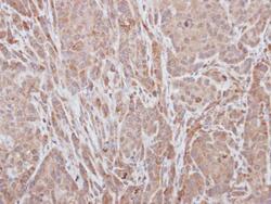
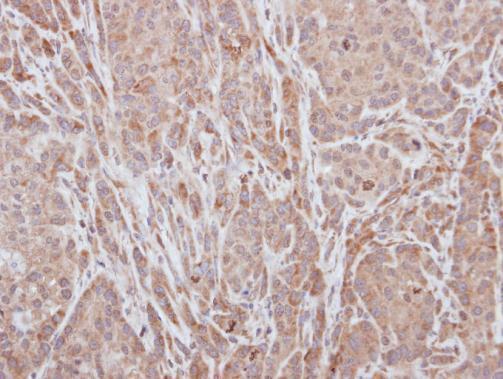
- Immunohistochemical analysis of paraffin-embedded A549 xenograft, using ADAMTS5 (Product # PA5-27165) antibody at 1:100 dilution. Antigen Retrieval: EDTA based buffer, pH 8.0, 15 min.
Supportive validation
- Submitted by
- Invitrogen Antibodies (provider)
- Main image
- Experimental details
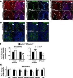
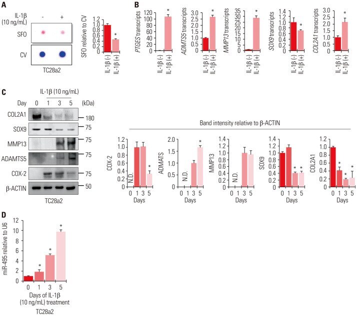
- Figure 7 Diminished ADAMTS and reduced versican cleavage in Galnt1 s. (A) ADAMTS1, expressed in the myocardium and developing cardiac cushion tissue (white dashed lines), is decreased at E12.5 in Galnt1 -/- relative to wild type ( Galnt1 +/+ ). (B) ADAMTS5, expressed in the myocardium and hinge region of developing valve leaflets (white dashed lines), is diminished at E12.5 and E14.5 in Galnt1 -/- relative to wild type ( Galnt1 +/+ ). (C) ADAMTS20, expressed in the myocardium and developing cardiac cushion tissues (white dashed lines), is unchanged at E12.5 in Galnt1 -/- . (D) Confocal images of intact versican (versican, green), (E) cleaved versican (DPEAAE; green), and nuclei (Nu; blue) show an increase in intact versican accompanied by a decrease in cleaved versican in E12.5 OFT cushions and E14.5 AoV of Galnt1 -/- relative to wild type. (F) Fluorescent staining intensity of intact versican at E12.5 (n = 5) and E14.5 (n = 5) and cleaved (DPEAAE) versican at E12.5 (n = 4) and E14.5 (n = 4) relative to nuclei staining, and normalized to wild type (equal to 1), at E12.5 and E14.5 shows significant increase in Galnt1 -/- OFT cushion tissue. (G) qPCR analysis of expression of major Adamts genes and versican ( Vcan ) from E12.5 OFT cushion (isolated by LCM) is unchanged in Galnt1 s relative to wild type (triplicates, n = 4). ***, P < 0.001. AoV, aortic valve. OFTC, outflow tract cushion. Scale bars: A, B, C, D, E = 10 mum.

- Submitted by
- Invitrogen Antibodies (provider)
- Main image
- Experimental details
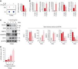
- Fig. 1 miRNA-495 is upregulated during osteoarthritis progression in an in vitro model generated using the human normal chondrocyte cell line TC28a2. (A) TC28a2 cells were seeded at 1x10 5 cells per well in 24-well culture plates. The cells were treated with 10 ng/mL of IL-1beta or with no IL-1beta as a control. Safranin O staining was performed to detect glycosaminoglycans (GAGs). Stained cells were destained with 10% cetylpyridinium for quantitative analysis. Absorbance was measured at 490 nm. * p
- Submitted by
- Invitrogen Antibodies (provider)
- Main image
- Experimental details
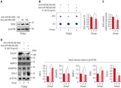
- Fig. 2 Inhibition of miRNA-495 expression during in vitro osteoarthritis progression protects chondrocytes from IL-1beta-mediated disruption of SOX9 and COL2A1. (A) Protein levels of SOX9 were analyzed in TC28a2 cells transfected with anti-miR negative control (NC, 100 nM), anti-miR-495 (50 or 100 nM) by Western blotting to determine the effective dose of anti-miR-495. (B) TC28a2 cells transfected with anti-miR NC (50 nM) or anti-miR-495 (50 nM) were seeded at 1x10 5 cells per well in 24-well culture plates. The cells were treated with 10 ng/mL of IL-1beta or with no IL-1beta as a control. Safranin O staining was performed to detect glycosaminoglycans (GAGs). Stained cells were destained with 10% cetylpyridinium for quantitative analysis. Absorbance was measured at 490 nm. * p

- Submitted by
- Invitrogen Antibodies (provider)
- Main image

- Experimental details
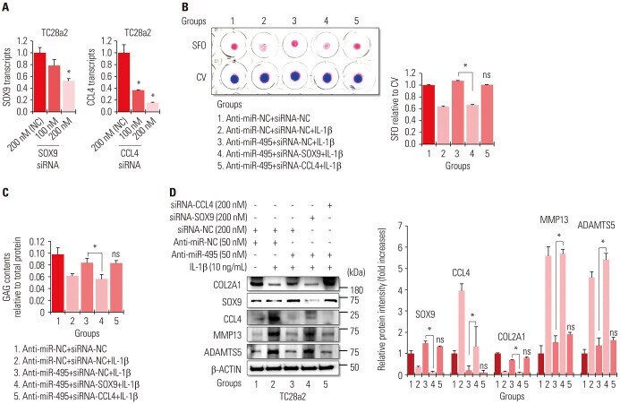
- Fig. 4 miRNA-495-SOX9 axis is more important than miRNA-495-CCL4 axis in protecting chondrocytes against IL-1beta-mediated inflammation. (A) The graphs represent the results of quantitative real-time polymerase chain reaction (qRT-PCR) using RNA extracted from TC28a2 cells transfected with negative control (NC) siRNA (200 nM), SOX9 siRNA (100 or 200 nM), or CCL4 siRNA (100 or 200 nM) (n=3 experimental replicates) to validate the efficiency of the siRNAs used in this study. The expression levels of SOX9 and CCL4 mRNAs were normalized to those of 18S rRNA. The data are presented as means+-SD. * p