Explore
 Validate
 Learn
Learn Immunohistochemistry
Antibody data
- Antibody Data
- Antigen structure
- References [3]
- Comments [0]
- Validations
- Immunohistochemistry [1]
Submit
Validation data
Reference
Comment
Report error
- Product number
- 600-406-106 - Provider product page

- Provider
- Rockland Immunochemicals, Inc.
- Proper citation
- Rockland Cat#600-406-106, RRID:AB_217582
- Product name
- Anti-Collagen Type IV (RABBIT) Antibody Biotin Conjugated - 600-406-106
- Antibody type
- Polyclonal
- Vial size
- 100 µl
Submitted references Magnetic Resonance Imaging Reveals Distinct Roles for Tissue Transglutaminase and Factor XIII in Maternal Angiogenesis During Early Mouse Pregnancy.
A developmentally regulated switch from stem cells to dedifferentiation for limb muscle regeneration in newts.
DNA Methylation Dynamics Regulate the Formation of a Regenerative Wound Epithelium during Axolotl Limb Regeneration.
Cohen G, Hadas R, Stefania R, Pagoto A, Ben-Dor S, Kohen F, Longo D, Elbaz M, Dekel N, Gershon E, Aime S, Neeman M
Arteriosclerosis, thrombosis, and vascular biology 2019 Aug;39(8):1602-1613
Arteriosclerosis, thrombosis, and vascular biology 2019 Aug;39(8):1602-1613
A developmentally regulated switch from stem cells to dedifferentiation for limb muscle regeneration in newts.
Tanaka HV, Ng NCY, Yang Yu Z, Casco-Robles MM, Maruo F, Tsonis PA, Chiba C
Nature communications 2016 Mar 30;7:11069
Nature communications 2016 Mar 30;7:11069
DNA Methylation Dynamics Regulate the Formation of a Regenerative Wound Epithelium during Axolotl Limb Regeneration.
Aguilar C, Gardiner DM
PloS one 2015;10(8):e0134791
PloS one 2015;10(8):e0134791
No comments: Submit comment
Supportive validation
- Submitted by
- Rockland Immunochemicals, Inc. (provider)
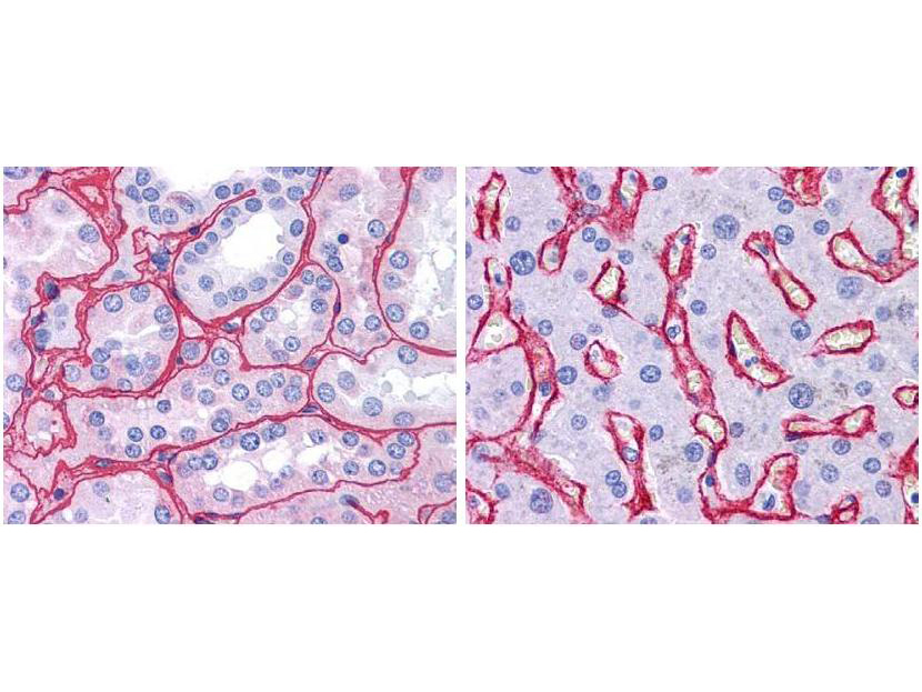
- Main image
- Experimental details
- Immunohistochemistry of Rabbit Anti-Collagen IV Biotin Conjugated Antibody. Tissue: human kidney (Left) with strong red staining observed in glomeruli and liver (Right) with strong staining in sinusoids. Fixation: formalin fixed paraffin embedded. Antigen retrieval: steamed in 0.01 M sodium citrate buffer, pH 6.0 at 99-100°C - 20 minutes. Primary antibody: Collagen IV antibody at 10 µg/mL for 1 h at RT. Secondary antibody: Peroxidase rabbit secondary antibody at 1:10,000 for 45 min at RT. Localization: Collagen IV is extracellular. Staining: Collagen IV as precipitated red signal with hematoxylin purple nuclear counterstain.
- Validation comment
- Immunohistochemistry