Explore
 Validate
 Learn
Learn Immunocytochemistry
Antibody data
- Antibody Data
- Antigen structure
- References [3]
- Comments [0]
- Validations
- Immunocytochemistry [1]
Submit
Validation data
Reference
Comment
Report error
- Product number
- 210-401-319 - Provider product page

- Provider
- Rockland Immunochemicals, Inc.
- Proper citation
- Rockland Cat#210-401-319, RRID:AB_10704513
- Product name
- Anti-Mouse IL-1ß (RABBIT) Antibody - 210-401-319
- Antibody type
- Polyclonal
- Vial size
- 100 µl
Submitted references Correction to: Niacin-mediated rejuvenation of macrophage/microglia enhances remyelination of the aging central nervous system.
Niacin-mediated rejuvenation of macrophage/microglia enhances remyelination of the aging central nervous system.
High Resolution Dissection of Reactive Glial Nets in Alzheimer's Disease.
Rawji KS, Young AMH, Ghosh T, Michaels NJ, Mirzaei R, Kappen J, Kolehmainen KL, Alaeiilkhchi N, Lozinski B, Mishra MK, Pu A, Tang W, Zein S, Kaushik DK, Keough MB, Plemel JR, Calvert F, Knights AJ, Gaffney DJ, Tetzlaff W, Franklin RJM, Yong VW
Acta neuropathologica 2020 May;139(5):911
Acta neuropathologica 2020 May;139(5):911
Niacin-mediated rejuvenation of macrophage/microglia enhances remyelination of the aging central nervous system.
Rawji KS, Young AMH, Ghosh T, Michaels NJ, Mirzaei R, Kappen J, Kolehmainen KL, Alaeiilkhchi N, Lozinski B, Mishra MK, Pu A, Tang W, Zein S, Kaushik DK, Keough MB, Plemel JR, Calvert F, Knights AJ, Gaffney DJ, Tetzlaff W, Franklin RJM, Yong VW
Acta neuropathologica 2020 May;139(5):893-909
Acta neuropathologica 2020 May;139(5):893-909
High Resolution Dissection of Reactive Glial Nets in Alzheimer's Disease.
Bouvier DS, Jones EV, Quesseveur G, Davoli MA, A Ferreira T, Quirion R, Mechawar N, Murai KK
Scientific reports 2016 Apr 19;6:24544
Scientific reports 2016 Apr 19;6:24544
No comments: Submit comment
Supportive validation
- Submitted by
- Rockland Immunochemicals, Inc. (provider)
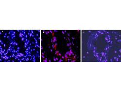
- Main image

- Experimental details
- Immunofluorescence microscopy after staining of mouse carotid artery tissue with anti-Mouse IL-1ß antiserum (less purified form of 210-401-319) diluted 1:50. Tissue sections were prepared after cyrofixation. Reaction occurred at room temperature for 60' followed by washes and reaction with Rhodamine conjugated Gt-a-Rabbit IgG (Rockland code 611-100-122). Tissue was counterstained with bis-benzimide solution at 0.5 mg/ml in PBS for 15 min at room temperature. Panel A) shows no antibody staining of WT uninjured mouse carotid tissue. Panel B) shows anti-IL-1ß staining of cells after surgical injury of tissue. Panel C) shows no antibody staining of injured carotid tissue from an IL-1ß KO mouse.
- Sample type
- IF Microscopy